
Abstract
Conclusion:
In peritoneal dialysis patients, residual renal function loss were associated with baseline sodium, triglyceride, and body mass index. There was no correlation between residual renal function loss and longitudinal CRP and uric acid levels. Prospective studies are needed to determine the optimal uric acid and CRP levels.
Results:
The follow-up period was 32.7 (12.9-36) months. Ten patients lost residual renal function 22.1±9.8 months after the initiation of peritoneal dialysis. Longitudinal uric acid level was 6.1±1.2 mg/dL and longitudinal CRP level was 0.5 (0.3-0.7) mg/dL. In patients with residual renal function loss, baseline sodium and triglyceride were lower, while parathormone were higher. There was no difference between the groups in terms of longitudinal uric acid and CRP levels. Baseline parathyroid hormone [hazard ratio (HR), 1,003; 95% confidence interval (CI) 1,001-1,006; p=0.013], body mass index (HR 0.817; 95% CI 0.684-0.975; p=0.025), and baseline sodium level (HR 0.801; 95% CI; 0.665- 0.965; p=0.019) were risk factors for residual renal function loss.
Materials and Methods:
This is a retrospective cohort study. Thirty-four patients who started peritoneal dialysis due to end-stage renal disease were included. The primary endpoint was loss of residual renal function and was defined as residual urine volume of less than 200 mL/24 hours. Patients were followed for three years after the onset of peritoneal dialysis or until loss of residual renal functions. Demographic and clinical data were recorded retrospectively.
Aim:
Residual renal functions have positive effects on morbidity and mortality among patients undergoing peritoneal dialysis. Our aim is to investigate the effects of baseline laboratory data and longitudinal uric acid and C-reactive protein (CRP) levels on loss of residual renal functions within the first three years of peritoneal dialysis.
Keywords: C-reactive protein, peritoneal dialysis, residual renal functions, sodium, uric acid
INTRODUCTION
Peritoneal dialysis (PD) is one of the renal replacement therapies in end-stage renal disease (ESRD). In patients undergoing PD, residual renal function (RRF) has positive effects on anemia, blood pressure and volume control, bone metabolism, and patient survival1-3. Therefore, it is important to maintain RRF in PD patients. Loss of RRF occurs earlier in patients with a history of diabetes mellitus (DM), heart failure, and peritonitis4. In addition, inflammation has also been found to be associated with loss of RRF5.
Uric acid is the end product of purine metabolism. It has been shown that uric acid causes endothelial dysfunction, vascular smooth muscle cell damage and inflammation6-8. Hyperuricemia is a risk factor for chronic kidney disease, diabetes and hypertension9-11. It has been reported that mortality is higher in hemodialysis and PD patients with high uric acid levels12,13. In epidemiological studies, it has been shown that the risk of chronic kidney disease increases in people with high uric acid levels11,14. In patients with chronic kidney disease, inconsistent results have been reported regarding the effect of uric acid on disease progression14-17. In two different studies examining the relationship between the basal uric acid level at the onset of PD and RRF, it was reported that high serum uric acid levels were associated with loss of RRF18,19. The number of studies examining the relationship between RRF loss and the longitudinal uric acid level measured until the loss of RRF and the loss of RRF is quite limited. In this study, it was aimed to investigate the effects of clinical and laboratory data before the initiation of PD and longitudinal uric acid and C-reactive protein (CRP) levels in the first three years after the initiation of PD on RRF.
GİRİŞ
Son dönem böbrek hastalığında (SDBH) böbrek yerine koyma tedavilerinden biri periton diyalizidir (PD). PD yapan hastalarda, rezidüel renal fonkisyonun (RRF), anemi, kan basıncı ve volüm kontrolü, kemik metabolizması ve hasta sağkalımı üzerinde olumlu etkileri vardır1-3. Bu nedenle, PD hastalarında RRF’nin korunması önemlidir. Diabetes mellitus (DM), kalp yetmezliği ve peritonit öyküsü olan hastalarda RRF kaybı daha erken olmaktadır4. Ayrıca, enflamasyon da RRF kaybı ile ilişkili bulunmuştur5.
Ürik asit, pürin metabolizmasının son ürünüdür. Ürik asidin, endotel disfonksiyonu, vasküler düz kas hücresi hasarı ve enflamasyona neden olduğu gösterilmiştir6-8. Hiperüriseminin, kronik böbrek hastalığı, diyabet ve hipertansiyon için risk faktörü olduğu rapor edilmiştir9-11. Hemodiyalize giren veya PD yapan hastalarda ise yüksek ürik asit düzeyine sahip olanlarda mortalitenin daha yüksek olduğu bildirilmiştir12,13. Epidemiyolojik çalışmalarda, ürik asit düzeyi yüksek olan kişilerde kronik böbrek hastalığı riskinin arttığı gösterilmiştir11,14. Kronik böbrek hastalığı olan kişilerde ise, ürik asidin hastalık progresyonu üzerindeki etkisine dair birbirinden farklı sonuçlar bildirilmiştir14-17. PD başlandığı andaki bazal ürik asit düzeyi ile RRF arasındaki ilişkinin incelendiği iki ayrı çalışmada, yüksek serum düzeyinin RRF kaybı ile ilişkili olduğu bildirilmiştir18,19. RRF kaybı olana kadar ölçülen boylamsal ürik asit düzeyinin RRF kaybı ile ilişkisinin incelendiği çalışma sayısı ise oldukça kısıtlıdır. Bu çalışmada, PD başlanmadan önceki klinik ve laboratuvar veriler ile PD başlandıktan sonraki ilk üç yıldaki boylamsal ürik asit ve C-reaktif protein (CRP) düzeyinin RRF üzerine olan etkisinin araştırılması amaçlanmıştır.
MATERIALS AND METHODS
In this retrospective cohort study, patients older than 18 years of age, who started PD due to ESRD between January 01, 2010 and October 31, 2018, were evaluated retrospectively. Exclusion criteria were: 1. Follow-up of less than three months after the initiation of PD (n=6), 2. Having a kidney transplant (n=12), and 3. Residual urine less than 200 mL/24 hours at the time of the initiation of PD and within three months (n=6). After excluding a total of 24 patients, 34 patients were included in our study. Ege University of Local Ethics Committee (protocol number: 21-11T/21, date: 04.11.2021) was obtained for our study. Patients underwent continuous ambulatory PD or automated PD.
In general, continuous ambulatory PD consisting of 4-5 exchanges/24 hours was applied to the patients. Patients who were treated with automated PD underwent six or more exchanges/24 hours. Dialysis type, dialysate and number of exchanges were determined according to the peritoneal equalization test, Kt/Vurea and ultrafiltration need of the patients. Dialysate glucose concentration and icodextrin requirement were adjusted according to the patient’s ultrafiltration need.
In our study, the primary endpoint was loss of RRF. RRF loss was defined as a residual urine volume of less than 200 mL/24 hours. Patients were followed for three years after the initiation of PD or until residual urine volume was <200 mL/24 hours. Patients who switched to another renal replacement therapy, died or were lost to follow-up were followed up to their last registered visit.
Demographic and clinical data (age, gender, height, weight, cause of ESRD, office blood pressure, DM, cardiovascular disease) and medications before the initiation of PD were recorded from patient files. Laboratory data (urea, creatinine, sodium, potassium, calcium, phosphorus, uric acid, albumin, CRP, triglyceride, low-density lipoprotein, parathormone (PTH), ferritin, hemoglobin) belonging to one month before starting PD were obtained from patient files and electronic hospital information management system. The type of PD at the initiation of PD and the weekly total Kt/Vurea measured between the third and sixth months after the initiation of PD were recorded. Body mass index (BMI) was obtained by dividing the patient’s body weight (kg) by the square of the height (m). Peritonitis attacks were also recorded. During the follow-up period, serum uric acid and CRP levels which were obtained during approximately quarterly visits were recorded from the patient files. If the patients had active infection during the visit, the CRP and uric acid levels at that visit were not included in the study. If the patient did not attend the visit, the CRP and uric acid levels evaluated within a month before or after that visit were recorded.
Statistical Analysis
Data analysis was performed with the statistical package program of IBM Statistical Package for the Social Sciences Statistics 25.0 (IBM Corp., Armonk, New York, USA). The distribution of normality for continuous variables was evaluated with the Shapiro-Wilk test. Descriptive statistics were given as frequency (n), percentage (%), mean, standard deviation, median (M), 25th percentile (C1), 75th percentile (C3). Longitudinal uric acid and CRP levels were calculated as the mean of uric acid and CRP levels measured during the follow-up period. Independent sample t-test, Mann-Whitney U test and Fisher’s exact chi-square test were used for comparisons between the groups. Risk factors for RRF loss were evaluated by cox regression analysis. A p value of <0.05 was considered statistically significant.
RESULTS
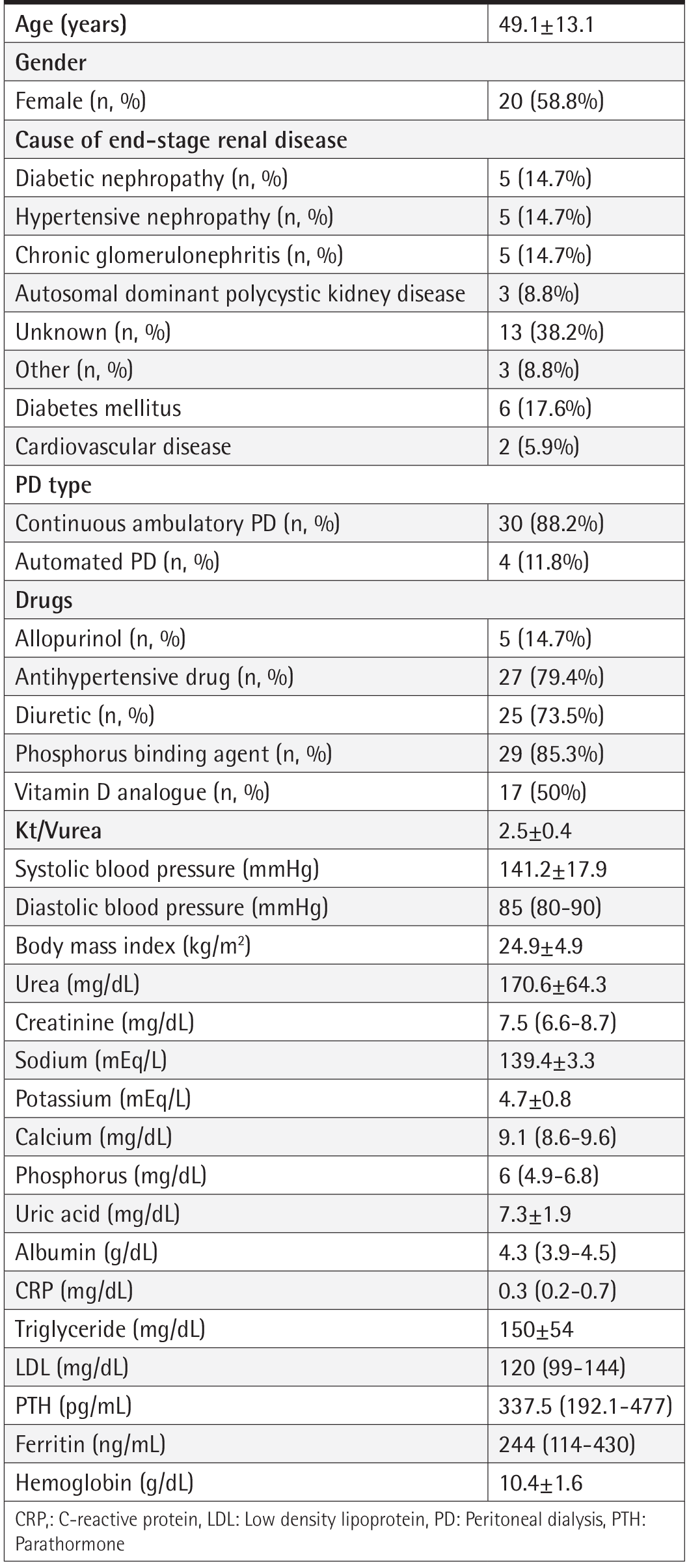
The study included 34 patients. Baseline demographic, clinical and laboratory data of the patients are shown in . The mean age of the patients was 49.1±13.1 years and 58.8% were female. The cause of ESRD was unknown in 38.2% of the patients. The most common causes of ESRD were diabetic nephropathy, hypertensive nephrosclerosis and chronic glomerulonephritis. Three patients had autosomal dominant polycystic kidney disease. At the time of initiation of PD, six patients had DM and two had cardiovascular disease. In tour study, 88.2% of the patients were undergoing continuous ambulatory PD. At the time of the initiation of PD, five patients were receiving allopurinol and 25 patients were receiving diuretics. Before the initiation of PD, baseline creatinine level was 7.5 (6.6-8.7) mg/dL, sodium level was 139.4±3.3 mEq/L, uric acid level was 7.3±1.9 mg/dL, and CRP level was 0.3 (0.2-0.7) mg/dL.
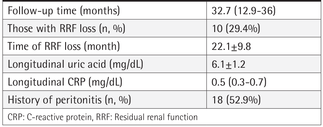
The mean follow-up period was 32.7 (12.9-36) months (). Longitudinal uric acid level was 6.1±1.2 mg/dL and longitudinal CRP level was 0.5 (0.3-0.7) mg/dL. The rate of patients who had peritonitis attack was 52.9%. RRF loss was observed in 10 patients 22.1±9.8 months after the initiation of PD.
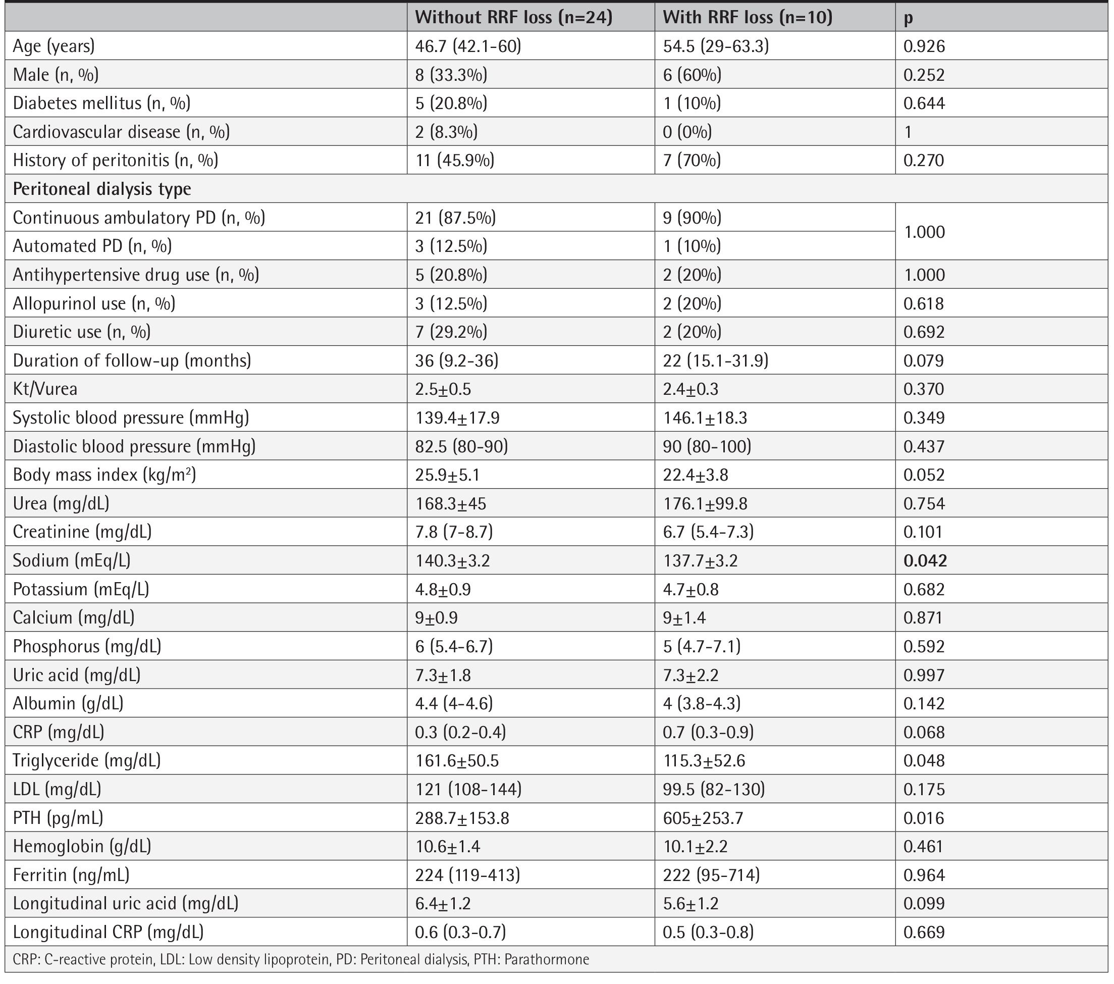
The baseline within data of patients with and without loss of RRF during the follow-up are shown in . There was no significant difference between the two groups in terms of age, gender, BMI and history of cardiovascular disease. Urea, creatinine, and hemoglobin levels were similar before the initiation of PD. Compared to patients without RRF loss, baseline sodium level was lower in those with RRF loss. Triglyceride was lower in those with RRF loss, while PTH was higher. BMI was lower in those with RRF loss, although it was not statistically significant. There was no difference between the two groups in terms of longitudinal uric acid and CRP levels.
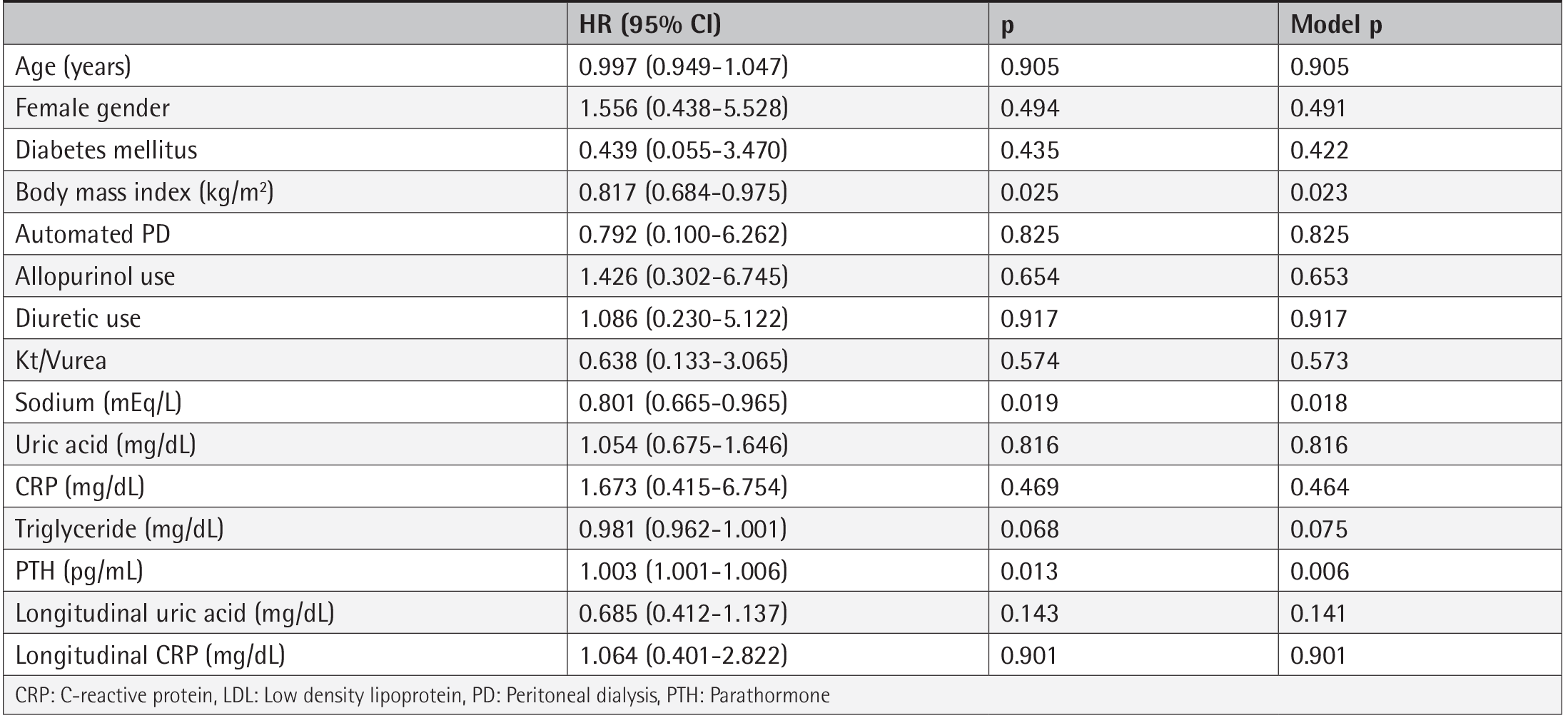
Baseline uric acid, baseline CRP, longitudinal uric acid and longitudinal CRP levels before the initiation of PD were not risk factors for loss of RRF (). The presence of DM, history of peritonitis and use of diuretics were also not associated with RRF loss. Before the initiation of PD, baseline sodium (HR 0.801; 95% CI 0.665-0.965, p=0.019), baseline PTH (HR, 1.003, 95% CI 1.001-1.006, p=0.013), and BMI (HR 0.817; 95%CI 0.684-0.975, p=0.025) were the risk factors for RRF loss. In the multivariate cox regression analysis including age at the initiation of PD, gender, DM, history of peritonitis, baseline PTH, baseline sodium, and baseline BMI, none of these parameters were risk factors for RRF loss.
DISCUSSION
In this study, 29.4% of the patients had loss of RRF 22.1±9.8 months after the initiation of PD. Compared to patients without RRF loss, patients with RRF loss had lower baseline sodium, lower triglyceride levels and higher PTH levels. Longitudinal uric acid and CRP levels were similar between the two groups. Baseline uric acid and CRP and longitudinal uric acid and CRP levels were not risk factors for RRF loss. Low sodium level, high PTH level, and low BMI at the initiation of PD were risk factors for RRF loss.
Loss of RRF develops in PD patients over time due to risk factors such as peritonitis, DM, and heart failure. In PD patients, RRF was found to be associated with better volume and blood pressure control, improvement of anemia, phosphorus control, and better survival1-3. Therefore, long-term preservation of RRF is sought in PD patients.
In our study, patients with RRF loss had lower sodium and triglyceride levels. The BMI and albumin levels of these patients were also lower, although not statistically significant. These laboratory findings are parameters related to nutrition. In addition, low triglyceride and sodium levels were found to be risk factors for RRF loss. Our findings support that RRF loss may be associated with malnutrition. In one study, malnutrition-inflammation complex syndrome was found to be associated with loss of RRF20. Palomo-Piñón et al.21 showed that baseline CRP level is high and albumin level is low in patients with RRF loss, and baseline CRP and albumin levels are risk factors for RRF loss. However, in our study, no correlation was found between baseline and longitudinal CRP levels and RRF.
Hyperuricemia causes inflammation, endothelial dysfunction and oxidative stress22. As hyperuricemia is associated with inflammatory markers such as interleukin-6, CRP, and tumor necrosis factor-alpha, it has been suggested that uric acid has a role in inflammatory processes8. In the general population, it has been shown that hyperuricemia is associated with hypertension, cardiovascular diseases and mortality23,24. In the CKD process, the results of studies on its relationship with disease progression and patient survival are contradictory14-17.
The number of studies examining the relationship between uric acid level and clinical outcomes in PD patients is very few. Park et al.18 Showed that there was a correlation between baseline uric acid level and loss of RRF at the end of 24-month follow-up. In a more recent study, a U-shaped relationship was found between baseline uric acid level and loss of RRF19. Most of the studies in the literature have evaluated the relationship between uric acid level at the initiation of PD and loss of RRF. In the only study examining the relationship between longitudinal uric acid levels and RRF after the initiation of PD, it was reported that longitudinal uric acid levels of <6.77 mg/dL and ≥7.64 mg/dL were associated with loss of RRF25. In our study, however, no relationship could be demonstrated between longitudinal uric acid level and loss of RRF. In our study, a relationship between RRF loss and longitudinal uric acid level may not have been determined due to the lower mean uric acid level and the small number of patients.
Study Limitations
The limitations of our study are its retrospective nature and the small number of patients. On the other hand, the longitudinal evaluation of uric acid and CRP levels can be considered as the strengths of our study.
TARTIŞMA
Bu çalışmada, hastaların %29,4’ünde PD başlandıktan 22,1±9,8 ay sonra RRF kaybı olduğu görüldü. RRF kaybı olmayan hastalar ile karşılaştırıldığında, RRF kaybı olan hastaların bazal sodyum, trigliserid düzeyi daha düşük ve PTH düzeyi daha yüksekti. Her iki grup arasında boylamsal ürik asit ve CRP düzeyleri benzer bulundu. Bazal ürik asit ve CRP ile boylamsal ürik asit ve CRP düzeyleri RRF kaybı için bir risk faktörü değildi. PD başlanmadan önceki sodyum düzeyinin düşük, PTH düzeyinin yüksek ve VKİ’nin düşük olması RRF kaybı için risk faktörü idi.
PD hastalarında zamanla böbrek işlevlerinin peritonit, DM, kalp yetmezliği gibi risk faktörleri nedeni ile RRF kaybı olur. PD hastalarında RRF daha iyi volüm ve kan basıncı kontrolü, aneminin iyileşmesi, fosfor kontrolü ve daha iyi sağkalım ile ilişkili bulunmuştur1-3. Bu nedenle, PD hastalarında RRF’nin uzun süre korunması sağlanmaya çalışılmaktadır.
Çalışmamızda, RRF kaybı olan hastaların sodyum ve trigliserid düzeyi daha düşüktü. Bu hastaların VKİ’leri ve albümin düzeyi de istatistiksel olarak anlamlı olmamakla birlikte daha düşüktü. Bu laboratuvar bulguları nütrisyon ile ilişkili parametrelerdir. Ayrıca, trigiliserid ve sodyum düşüklüğü RRF kaybı için risk faktörü olarak bulunmuştur. Bulgularımız, RRF kaybının malnütrisyon ile ilişkili olabileceğini desteklemektedir. Bir çalışmada, malnütrisyon-enflamasyon kompleksi sendromu RRF kaybı ile ilişkili bulunmuştur20. Palomo-Piñón ve ark.21 RRF kaybı olan hastalarda bazal CRP düzeyinin yüksek, albümin düzeyinin düşük olduğunu ve bazal CRP ve albümin düzeyinin RRF kaybı için risk faktörü olduğunu göstermişlerdir. Fakat, çalışmamızda bazal ve boylamsal CRP düzeyi ile RRF arasında bir ilişki bulunmamıştır.
Hiperüriseminin enflamasyona, endotel disfonksiyonuna ve oksidatif strese neden olduğu bilinmektedir22. Hiperüriseminin interlökin-6, CRP ve tümor nekrozis faktör-alfa gibi enflamasyon belirteçleri ile ilişkili olduğu, dolayısıyla enflamatuvar süreçlerde rol aldığı öne sürülmüştür8. Genel popülasyonda, hiperüriseminin hipertansiyon, kardiyovasküler hastalıklar ve mortalite ile ilişkili olduğu gösterilmiştir23,24. KBH sürecinde ise, hastalık progresyonu ve hasta sağkalımı ile ilişkisine dair çalışmaların sonuçları çelişkilidir14-17.
PD hastalarında ürik asit düzeyi ile klinik sonlanımların ilişkisini inceleyen çalışma sayısı oldukça azdır. Park ve ark.18, 24 aylık takip sonucunda bazal ürik asit düzeyi ile RRF kaybı arasında ilişki olduğunu göstermişlerdir. Daha güncel bir çalışmada ise bazal ürik asit düzeyi ile RRF kaybı arasında U-şeklinde bir ilişki saptanmıştır19. Literatürdeki çalışmaların çoğu, PD başlandığı sıradaki ürik asit düzeyi ile uzun vadede gelişen RRF kaybı arasındaki ilişkiyi incelemişlerdir. PD başlandıktan sonraki boylamsal ürik asit düzeylerinin RRF ile ilişkisini inceleyen tek çalışmada, boylamsal ürik asit düzeyinin <6,77 mg/dL ve ≥7,64 mg/dL olmasının RRF kaybı ile ilişkili olduğu bildirilmiştir25. Bizim çalışmamızda ise, boylamsal ürik asit düzeyi ile RRF kaybı arasında bir ilişki gösterilememiştir. Çalışmamızda, ortalama ürik asit düzeyinin daha düşük olması ve hasta sayısının az olması nedeni ile RRF kaybı ve boylamsal ürik asit düzeyi arasında bir ilişki saptanamamış olabilir.
Çalışmanın Kısıtlılıkları
Çalışmamızın kısıtlılıkları retrospektif olması ve hasta sayısının az olmasıdır. Bununla birlikte, ürik asit ve CRP düzeylerinin boylamsal olarak değerlendirilmiş olması ise çalışmamızın güçlü yönü olarak kabul edilebilir.
CONCLUSION
In conclusion, while loss of RRF was associated with low baseline sodium, triglyceride and BMI in PD patients, no relationship could be shown between longitudinal CRP and uric acid levels and RRF loss. Larger, prospective studies are needed to determine the effects of hyperuricemia on RRF loss and, in this respect, to determine the target uric acid level.
Footnotes
Ethics
Ethics Committee Approval: The study were approved by the Ege University of Local Ethics Committee (protocol number: 21-11T/21, date: 04.11.2021).
Informed Consent: It is a retrospective cohort study.
Peer-review: Externally peer-reviewed.
Authorship Contributions
Concept: A.Ç., M.S.D., M.Ö., Design: A.Ç., M.Y., M.S.D., G.A., H.T., M.Ö., Data Collection or Processing: A.Ç., Z.A., M.Y., M.S.D., G.A., H.T., M.Ö., Analysis or Interpretation: A.Ç., Z.A., M.Y., G.A., H.T., M.Ö., Literature Search: A.Ç., Z.A., M.Y., Writing: A.Ç., M.Y., M.S.D., H.T., M.Ö.
Conflict of Interest: No conflict of interest was declared by the authors.
Financial Disclosure: The authors declared that this study received no financial support.
References
López-Menchero R, Miguel A, García-Ramón R, Pérez-Contreras J, Girbés V. Importance of residual renal function in continuous ambulatory peritoneal dialysis: its influence on different parameters of renal replacement treatment. Nephron. 1999;83:219-25.
Perl J, Bargman JM. The importance of residual kidney function for patients on dialysis: a critical review. Am J Kidney Dis. 2009;53:1068-81.
Paniagua R, Amato D, Vonesh E, Correa-Rotter R, Ramos A, Moran J, et al. Effects of increased peritoneal clearances on mortality rates in peritoneal dialysis: ADEMEX, a prospective, randomized, controlled trial. J Am Soc Nephrol. 2002;13:1307-20.
Marrón B, Remón C, Pérez-Fontán M, Quirós P, Ortíz A. Benefits of preserving residual renal function in peritoneal dialysis. Kidney Int Suppl. 2008;(108):S42-51.
Chung SH, Heimbürger O, Stenvinkel P, Qureshi AR, Lindholm B. Association between residual renal function, inflammation and patient survival in new peritoneal dialysis patients. Nephrol Dial Transplant. 2003;18:590-7.
Khosla UM, Zharikov S, Finch JL, Nakagawa T, Roncal C, Mu W, et al. Hyperuricemia induces endothelial dysfunction. Kidney Int. 2005;67:1739-42.
Mazzali M, Kanellis J, Han L, Feng L, Xia YY, Chen Q, et al. Hyperuricemia induces a primary renal arteriolopathy in rats by a blood pressure-independent mechanism. Am J Physiol Renal Physiol. 2002;282:F991-7.
Lyngdoh T, Marques-Vidal P, Paccaud F, Preisig M, Waeber G, Bochud M, et al. Elevated serum uric acid is associated with high circulating inflammatory cytokines in the population-based Colaus study. PLoS One. 2011;6:e19901.
Han T, Meng X, Shan R, Zi T, Li Y, Ma H, et al. Temporal relationship between hyperuricemia and obesity, and its association with future risk of type 2 diabetes. Int J Obes (Lond). 2018;42:1336-44.
Wang J, Qin T, Chen J, Li Y, Wang L, Huang H, et al. Hyperuricemia and risk of incident hypertension: a systematic review and meta-analysis of observational studies. PLoS One. 2014;9:e114259.
Ryoo JH, Choi JM, Oh CM, Kim MG. The association between uric acid and chronic kidney disease in Korean men: a 4-year follow-up study. J Korean Med Sci. 2013;28:855-60.
Xiang S, Zhang X, Xie X, Wang J, Zhou Q, Chen Z, et al. High serum uric acid level is a mortality risk factor in peritoneal dialysis patients: a retrospective cohort study. Nutr Metab (Lond). 2019;16:52.
Jeon JS, Chung SH, Han DC, Noh H, Kwon SH, Lindholm B, Lee HB. Mortality predictive role of serum uric acid in diabetic hemodialysis patients. J Ren Nutr. 2014;24:336-42.
De Cosmo S, Viazzi F, Pacilli A, Giorda C, Ceriello A, Gentile S, et al. Serum Uric Acid and Risk of CKD in Type 2 Diabetes. Clin J Am Soc Nephrol. 2015;10:1921-9.
Madero M, Sarnak MJ, Wang X, Greene T, Beck GJ, Kusek JW, et al. Uric acid and long-term outcomes in CKD. Am J Kidney Dis. 2009;53:796-803.
Srivastava A, Kaze AD, McMullan CJ, Isakova T, Waikar SS. Uric Acid and the Risks of Kidney Failure and Death in Individuals With CKD. Am J Kidney Dis. 2018;71:362-70.
Tsai CW, Chiu HT, Huang HC, Ting IW, Yeh HC, Kuo CC. Uric acid predicts adverse outcomes in chronic kidney disease: a novel insight from trajectory analyses. Nephrol Dial Transplant. 2018;33:231-41.
Park JT, Kim DK, Chang TI, Kim HW, Chang JH, Park SY, et al. Uric acid is associated with the rate of residual renal function decline in peritoneal dialysis patients. Nephrol Dial Transplant. 2009;24:3520-5.
Hsieh YP, Yang Y, Chang CC, Kor CT, Wen YK, Chiu PF, et al. U-shaped relationship between uric acid and residual renal function decline in continuous ambulatory peritoneal dialysis patients. Nephrology (Carlton). 2017;22:427-35.
Szeto CC, Kwan BC, Chow KM, Chung S, Yu V, Cheng PM, et al. Predictors of residual renal function decline in patients undergoing continuous ambulatory peritoneal dialysis. Perit Dial Int. 2015;35:180-8.
Palomo-Piñón S, Mora-Villalpando CJ, Del Carmen Prado-Uribe M, Ceballos-Reyes GM, De Jesús Ventura-García M, Ávila-Díaz M, et al. Inflammation and myocardial damage markers influence loss of residual renal function in peritoneal dialysis patients. Arch Med Res. 2014;45:484-8.
Filiopoulos V, Hadjiyannakos D, Vlassopoulos D. New insights into uric acid effects on the progression and prognosis of chronic kidney disease. Ren Fail. 2012;34:510-20.
Fang J, Alderman MH. Serum uric acid and cardiovascular mortality the NHANES I epidemiologic follow-up study, 1971-1992. National Health and Nutrition Examination Survey. JAMA. 2000;283:2404-10.
Sundström J, Sullivan L, D’Agostino RB, Levy D, Kannel WB, Vasan RS. Relations of serum uric acid to longitudinal blood pressure tracking and hypertension incidence. Hypertension. 2005;45:28-33.
Yang C, Ma X, Zhao W, Chen Y, Lin H, Luo D, ey al. A longitudinal analysis of the relationship between serum uric acid and residual renal function loss in peritoneal dialysis patients. Ren Fail. 2020;42:447-54.