
Abstract
Toxoplasmosis is an infectious disease caused by a protozoan parasite called Toxoplasma gondii. Rarely, undiagnosed Human Immunodeficiency virus patients may be diagnosed with cerebral toxoplasmosis as the etiology of confusion and headache. Our case is a 26-year-old man who presented with a disturbance of consciousness and headache for a week and was admitted to the hospital. There were no significant features in the patient’s medical and family history. Magnetic resonance imaging and CD4 positive T-cell blood level revealed a high suspicion of toxoplasma infection. Polymerase chain reaction test for Toxoplasma gondii deoxyribonucleic acid in his cerebrospinal fluid was positive. The patient was diagnosed with cerebral toxoplasmosis and acquired immunodeficiency syndrome. Trimethoprim-sulfamethoxazole (TMP-SMX) was initiated at 1600 mg SMX/320 mg TMP two times a day and clindamycin 900 mg three times a day. Tenofovir, emtricitabine and efavirenz were added. His symptoms gradually improved over 72 hours and after 21 days, the patient was discharged from the hospital. While toxoplasmosis is often curable, it poses a serious health threat to immunocompromised individuals and can be fatal if not promptly treated.
Keywords: Cerebral toxoplasmosis, confusion, headache, neuroinfection
INTRODUCTION
Toxoplasmosis is an infectious disease caused by a protozoan parasite called Toxoplasma gondii. It occurs by consuming contaminated food sources such as water, undercooked meat, and cat feces. Severe toxoplasmosis causing damage to the brain is most likely in immunocompromised patients such as those with Human Immunodeficiency Virus (HIV). The incidence of toxoplasmosis in HIV-infected patients increases with a CD4+ count below 200 cells/μL. The greatest risk is in patients with CD4+ counts less than 50 cells/μL. Central nervous system (CNS) toxoplasmosis rarely results from primary infection. Seroprevalence of anti-toxoplasma antibody varies substantially among different geographic locales, with a prevalence of approximately 11% in the United States, versus 50% to 80% in certain European, Latin American, and African countries. In toxoplasmosis seroprevalence studies conducted in Türkiye, it was reported that IgG antibody positivity was 28.8% in pregnant women, 31.9% in the population with HIV infection and even 65.2% in women living in rural areas. Cerebral toxoplasmosis is observed in 10-34% of autopsies conducted on patients with HIV. Common symptoms of toxoplasma encephalitis are flu-like symptoms, headache, seizures, hemiparesis, and mental disorders. Extracerebral involvement can also be seen including but not limited to pneumonitis, chorioretinitis, etc., .
CASE REPORT
Our case is a 26-year-old man of Turkish ethnicity, who presented with a disturbance of consciousness and headache for a week and was admitted to the hospital. There were no significant features in the patient’s medical and family history. Written informed consent was obtained from the patient to present this case. This study was performed in line with the principles of the Declaration of Helsinki.
Patient’s systemic examination was normal. Neurological examination revealed apathy, somnolence, bilateral positive Babinski’s sign, and increased deep tendon reflexes. Magnetic resonance imaging (MRI) revealed a high index of suspicion of a toxoplasma infection (). His CD4+ count was 87 cells/μL and his HIV1-RNA viral load was 6.2 × 10⁵ copies/mL. The serum Toxoplasma gondii IgG antibody measured with enzyme-linked immunosorbent assay was over 250 IU/mL (). Polymerase chain reaction test for Toxoplasma gondii DNA in his cerebrospinal fluid was positive. The patient was diagnosed with cerebral toxoplasmosis and Acquired Immunodeficiency syndrome (AIDS). Trimethoprim-sulphametoxazole (TMP-SMX) was initiated at 1600 mg SMX/320 mg TMP two times a day and clindamycin 900 mg three times a day. Tenofovir, emtricitabine, and efavirenz were started. His symptoms gradually improved over 72 hours and after 21 days, the patient was discharged from the hospital.
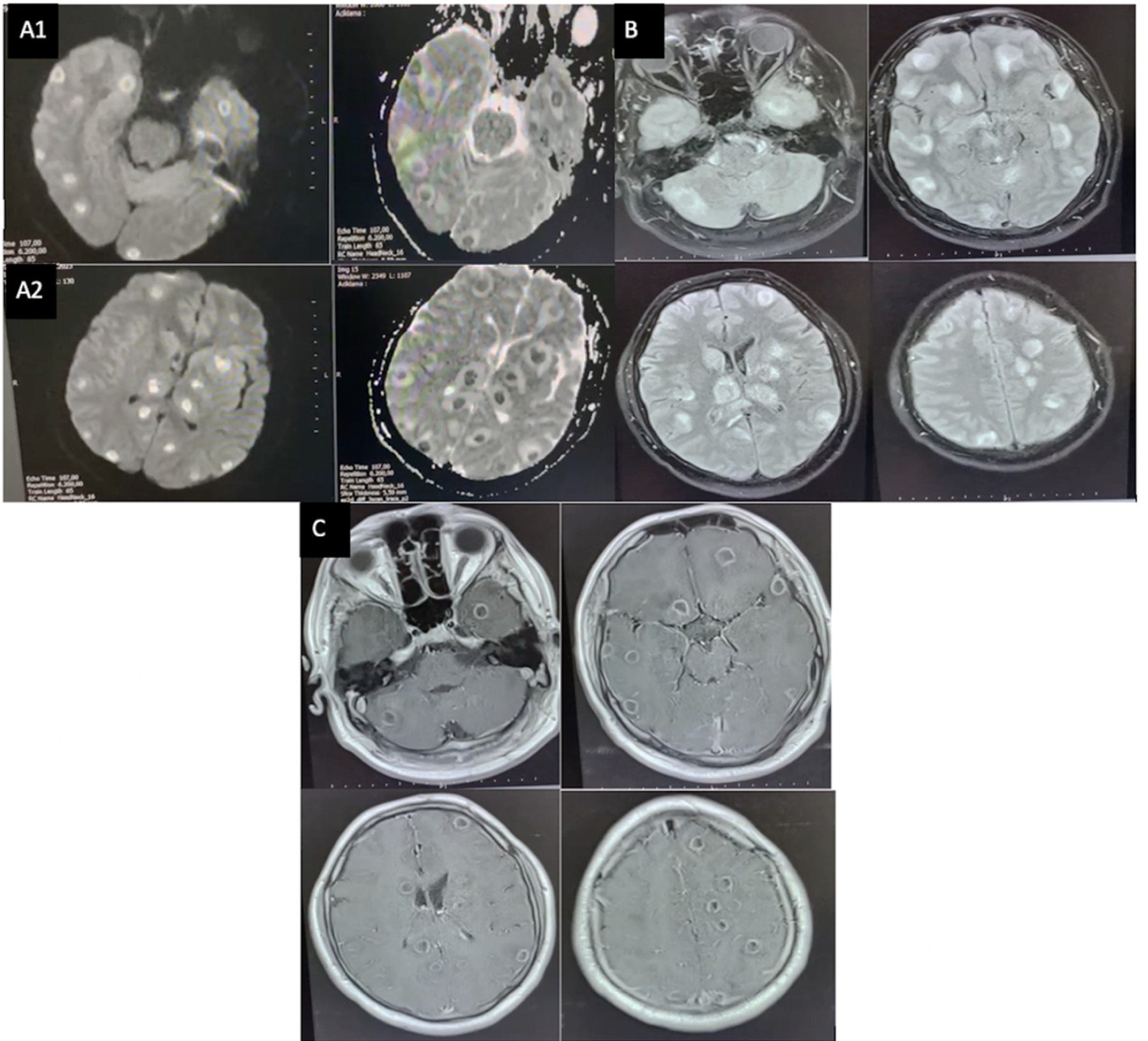
MRI: Magnetic resonance imaging
Table 1. Cerebrospinal fluid findings
Appearance | Clear |
Color | Colorless |
Cells | 10/mm3 leukocytes (lymphocytes) |
Total protein | 120 mg/dL |
Glucose | 63 mg/dL |
DISCUSSION
Toxoplasma gondii is the leading cause of brain lesions in patients with AIDS. Diagnosis of the cerebral toxoplasmosis requires the presence of three components; clinical symptoms corresponding to toxoplasma infection, specific CNS lesions detected by computed tomography or MRI scans and positive Toxoplasma gondii serological tests. All of these criteria are present in the described case. Considering differential diagnoses, cerebral cysticercosis is associated with epileptic seizures, headache, meningeal irritation, cognitive disorders, hemiparesis and palsy. Mononuclear pleocytosis, eosinophils, elevated levels of protein, and normal or lower levels of blood glucose are detected in the cerebrospinal fluid. The diagnosis is set on grounds of the ELISA, which is used for the detection of specific IgM antibodies. The changes demonstrated by the laboratory test results in our patient were mild proteinemia and pleocytosis with a high prevalence of the lymphocytes and normal glucose levels. In adult patients, tuberculosis (TB) of the CNS is secondary, developing after the primary TB infection located most often in the lungs. The imaging results reveal a large number of small-sized foci situated in the basal region of the brain. In the described case, lung imaging does not suggest TB. Multiple cerebral abscesses are associated with various infectious diseases (aspergillosis, cryptococcosis), neoplasms, and vasculitis. The imaging tests show a significant number of ring-like lesions similar to those observed in our case. However, clinically, brain abscesses are typically associated with persistent fever, recurrent seizures, visual disturbances, focal neurological deficits, and hemiparesis. However, our patient did not have fever. Multiple brain tumor metastases result from a primary cryptogenic malignant process. 40% of the gliomas and blastomas are demonstrated by similar ring-like lesions. In our case, we found no evidence of a primary neoplastic process. In multiple sclerosis, demyelinating lesions do not look like a closed ring. In our patient, the clinical criteria for this diagnosis were not present.
The standard treatment involves a combination of pyrimethamine, sulfadiazine, and folinic acid. TMP-SMX can be used as an alternative regimen. Clindamycin is an option for patients allergic to sulfa drugs. Effective antiretroviral therapy is equally important, . In our study, treatment success was achieved with the combination of TMP-SMX and clindamycin. With antibiotic therapy, 74% of patients improve by day 7 and 91% improve by day 14. Imaging studies are performed every 4-6 weeks until the complete resolution of the lesion or stabilization after partial resolution. The primary therapy is administered for 6 weeks, after which long-term suppressive therapy at lower doses is continued, with the duration based on the response to Highly Active Antiretroviral Therapy (HAART). HAART typically involves a combination of three or more antiretroviral drugs. The most common treatment combination is two nucleoside reverse transcriptase inhibitors (tenofovir-emtricitabine) plus a non-nucleoside reverse transcriptase inhibitor or integrase strand transfer inhibitor. In our case, tenofovir, emtricitabine, and efavirenz were started. The long-term suppressive therapy can be discontinued in patients with persistent elevation of CD4+ counts greater than 200 cells/μL and resolution of lesions on MRI.
To prevent primary toxoplasmosis, patients should refrain from consuming undercooked meat and ensure thorough handwashing after handling soil or coming into contact with cats. Patients who are seropositive for toxoplasma should be started on prophylaxis against CNS toxoplasmosis if their CD4+ count drops below 100 cells/μL.
The use of antiretroviral therapy markedly decreases the incidence of cerebral toxoplasmosis. Nevertheless, this disease continues to be the leading cause of expansive brain lesions, resulting in high morbidity and mortality among individuals with advanced immunosuppression, especially in low- and middle-income countries. Anti-toxoplasma therapy is an important component of the diagnostic approach to expansive brain lesions in AIDS. Local neuroepidemiology, degree of immunosuppression, and individual clinical, laboratory and neuroradiological features are extremely important in making differential diagnoses. TMP-SMX can be used for primary prophylaxis, initial therapy, and secondary prophylaxis of HIV-associated cerebral toxoplasmosis. Early initiation of antiretroviral therapy is currently possible because more effective and safer treatment options are available.
CONCLUSION
Cerebral toxoplasmosis is a common and treatable condition in HIV-positive patients; therefore, we aimed to present our case. Due to logistical challenges, a follow-up brain MRI could not be performed on the patient presented in our article after clinical improvement. Only clinical improvement was observed, and the patient failed to adhere to follow-up appointments. The lack of follow-up imaging and the patient’s non-compliance with follow-up visits are the limitations of our article, as the clinical outcome of the case remains unknown. While toxoplasmosis is often curable, it poses a serious health threat to immunocompromised individuals and can be fatal if not promptly treated.
Footnotes
Ethics
Informed Consent: Written informed consent was obtained from the patient to present this case. This study was performed in line with the principles of the Declaration of Helsinki.
Authorship Contributions
Surgical and Medical Practices: İ.K., Concept: İ.K., Design: İ.K., T.K.A., Data Collection or Processing: İ.K., Analysis or Interpretation: İ.K., Literature Search: İ.K., T.K.A, Writing: İ.K., T.K.A.
Conflict of Interest: No conflict of interest was declared by the authors.
Financial Disclosure: The authors declared that this study received no financial support.
References
Cook AJ, Gilbert RE, Buffolano W, Zufferey J, Petersen E, Jenum PA, et al. Sources of toxoplasma infection in pregnant women: European multicentre case-control study. European Research Network on Congenital Toxoplasmosis. BMJ. 2000;321:142-7.
Bowen LN, Smith B, Reich D, Quezado M, Nath A. HIV-associated opportunistic CNS infections: pathophysiology, diagnosis and treatment. Nat Rev Neurol. 2016;12:662-74.
Kaplan JE, Benson C, Holmes KH, Brooks JT, Pau A, Masur H. Guidelines for prevention and treatment of opportunistic infections in HIV-infected adults and adolescents: recommendations from CDC, the National Institutes of Health, and the HIV Medicine Association of the Infectious Diseases Society of America. MMWR Recomm Rep. 2009;58:1-207.
Carme B, Bissuel F, Ajzenberg D, Bouyne R, Aznar C, Demar M, et al. Severe acquired toxoplasmosis in immunocompetent adult patients in French Guiana. J Clin Microbiol. 2002;40:4037-44.
National Institutes of Health. Guidelines for the Prevention and Treatment of Opportunistic Infections in Adults and Adolescents with HIV: Toxoplasma gondii Encephalitis. Available from: https://clinicalinfo.hiv.gov/en/guidelines/hiv-clinical-guidelines-adult-and-adolescent-opportunistic-infections/toxoplasmosis?guideline%5B0%5D=title_bookpart%3AHIV+Clinical+Guidelines%3A+Adult+and+Adolescent+ Opportunistic+Infectio ns&redirected=1
Senoglu S, Yesilbag Z, Aydin OA, Karaosmanoglu HK, Yasar KK. Toxoplasma gondii IgG seroprevalence in patients with HIV/AIDS. Turkiye Parazitol Derg. 2018;42:175-9.
Kornienko VN, Pronin IN. Diagnostic neuroradiology. AJNR Am J Neuroradiol. 2009;30:E136.
Porter SB, Sande MA. Toxoplasmosis of the central nervous system in the acquired immunodeficiency syndrome. N Engl J Med. 1992;327:1643-8.
Pereira-Chioccola VL, Vidal JE, Su C. Toxoplasma gondii infection and cerebral toxoplasmosis in HIV-infected patients. Future Microbiol. 2009;4:1363-79.
Rothman RE, Marco CA, Yang S. Human immunodeficiency virus infection and acquired immunodeficiency syndrome in: Tintinalli JE, Stapczynski JS, Cline DM, Ma OJ, Cydulka RK, Meckler GD. Tintinalli’s emergency medicine: a comprehensive study guide. 7th ed. Chapter 149, New York; McGraw- Hill; 2011.
Kimura-Hayama ET, Higuera JA, Corona-Cedillo R, Chávez-Macías L, Perochena A, Quiroz-Rojas LY, et al. Neurocysticercosis: radiologic-pathologic correlation. Radiografics. 2010;30:1705-19
Khatri GD, Krishnan V, Antil N, Saigal G. Magnetic resonance imaging spectrum of intracranial tubercular lesions: one disease, many faces. Pol J Radiol. 2018;83:524-35.
Krishna K, Sada E, Vikram A, Gupta A. Multiple brain abscesses - diagnostic dilemma and therapeutic nightmare. J Neurosci Rural Pract. 2013;4:234-6.
Fink KR, Fink JR. Imaging of brain metastases. Surg Neurol Int. 2013;4:209-19.
Ge Y. Multiple sclerosis: the role of MR imaging. Am J Neuroradiol. 2006;27:1165-76.
Dedicoat M, Livesley N. Management of toxoplasmic encephalitis in HIV-infected adults (with an emphasis on resource-poor settings). Cochrane Database Syst Rev. 2006;19:5420.
Bertschy S, Opravil M, Cavassini M, Bernasconi E, Schiffer V, Schmid P, et al. Discontinuation of maintenance therapy against toxoplasma encephalitis in AIDS patients with sustained response to anti-retroviral therapy. Clin Microbiol Infect. 2006;12:666-71.
Eggleton JS, Nagalli S. Highly Active Antiretroviral Therapy (HAART). In: StatPearls. Treasure Island (FL): StatPearls Publishing; 2024.