
Abstract
Aim
Arteriovenous fistula (AVF) is a cannulation method that is accessed by a peripheric vein and an artery. AVF provides vascular access for chronic kidney disease patients so they can receive hemodialysis. AVF could be created by surgical intervention and facilitates arterial to venous circulation for rapid recovery. However, AVF maturation depends on venous proliferation and luminal diameter which allows the optimum flow rate for continuing circiulation and hemodialysis. Due to multiple unexpected conditions, non-maturation of AVFs limits the efficacy of the hemodialysis so patients must receive another surgery for AVF cannulation.
Materials and Methods
In this study, we aimed to utilize the effects of melatonin (MT), which is known to have antioxidant, anti-inflammatory, and antiapoptotic effects, to provide longer and more effective use of AVFs via a novel technique. For this purpose, firstly by electrospinning method, polylactic-co-glycolic acid (PLGA) nanofiber membranes were developed. After MT is loaded into the PLGA and characterized. Biodegradation and drug release profiles were analyzed. An in vivo study was performed in Wistar Albino male rats (n=18). Rats were randomly divided into three experimental groups; Sham, PLGA, and MT/PLGA respectively (n=6). AVF model was established in all groups between arteria carotica and vena jugularis under general anesthesia. The Sham group did not receive any treatment or biomaterial application. The developed membranes were placed onto the AVFs in PLGA and MT/PLGA groups. All rats were sacrificed on the 28th of the experiment. The anastomosis sites of all animals were harvested for histopathological analysis.
Results
Our results showed MT/PLGA group indicated increased maturation levels conmpared to Sham group (p<0.05).
Conclusion
The results showed that PLGA/MEL may be a promising material for early AVF maturation.
Keywords: Arteriovenous fistula, melatonin, PLGA, biocompatible materials, maturation, venous proliferati
INTRODUCTION
Chronic kidney disease (CKD) treatment in the early stages is primarily aimed at preserving the existing kidney function and includes methods such as diet, adequate fluid intake, medical treatments such as the use of angiotensin inhibitors, lifestyle changes, and increased physical activity. In advanced renal failure, hemodialysis and peritoneal dialysis are the main treatments applied, while kidney transplantation is the last resort for end-stage renal failure patients. According to the Global Diagnosis Burden 2017 data, CKD affects approximately 700 million people worldwide. Due to the extreme increase in the number of patients and the shortage of organ donors, CKD patients can only survive with hemodialysis. Hemodialysis is indispensable for CKD patients to balance minerals in circulating blood and control blood pressure. For long-term hemodialysis, patients need permanent vascular access established between arteries in a venous line, called an arteriovenous fistula (AVF). AVF is the most common cannulation method for long-term hemodialysis patients. Primary maturation failure is the main limiting factor for AVF use due to internal remodeling and lumen stenosis. Previous studies have claimed that vein luminal diameter below 2 mm is prone to deterioration. In addition, it is widely accepted that the vein diameter should be higher than the anastomotic artery diameter. Therefore, early maturation is urgent for long-term use of AVF. Otherwise, patients have to undergo several surgeries to have another AVF cannulation for hemodialysis treatment.
The immaturity of AVFs is mostly due to thickening of the vascular intima due to underlying pathological mechanisms consisting of oxidative stress, inflammation or hypoxia, which leads to abundant migration and proliferation of smooth muscle cells and accumulation of extracellular matrix. Studies have reported that AVF failure mostly occurs in the early weeks after surgery. At approximately 4 to 6 weeks, if blood flow is less than 500 mL/min. and the vessel diameter is less than 4 mm, urgent reoperation is required to reestablish a new AVF access. Therefore, establishing successful AVF cannulation as well as ensuring AVF maturity are essential for long-term hemodialysis. Despite the use of improved surgical techniques and new pharmacotherapy agents, there is no gold standard method to prevent AVF failure. Therefore, in this study, we aimed to prevent early maturation of AVF and avoid stenosis to prevent insufficient blood flow between artery and vein.
At this point, tissue engineering offers promising results for the treatment of various pathologies with its biocompatibility properties. Synthetic polymers are the most widely used carriers in the field of tissue engineering studies. Polylactic-co-glycolic acid (PLGA) is one of the preferred synthetic polymers for carrying various agents, cells or molecules. It consists of lactic acid and glycolic acid that can activate cell migration, proliferation and adhesion. Due to its biocompatibility and biodegradability, PLGA provides beneficial effects for tissue healing or regeneration. In our study, the effects of melatonin (MT) loaded PLGA nanofibers on AVF maturation were investigated. MT loaded PLGA biomaterials are frequently preferred in the field of tissue engineering. Since MT and PLGA are Food and Drug Administration (FDA)-approved molecules, it is stated that they are quite suitable for clinical translation-. In addition, the anti-inflammatory and antioxidant properties of MT provide a treatment-enhancing feature in the treatment of many pathogenesis. On the other hand, the feature of PLGA, which mimics the extracellular matrix of cells, strengthens this synergy and provides various positive results in experimental studies. MT is a hormone secreted from the pineal gland, with a hydrophilic structure that allows all cell compartments to show their effects. The antioxidant and anti-inflammatory effects of MT are well known, . In addition, MT has healing effects on the cardiovascular system by reducing iNOS and eNOS synthesis and increasing angiogenesis. Controlled release studies of MT have gained importance in recent years 15. The reasons for this may be that oral or parenteral use of MT limits its effect in the first pass through the liver and does not provide sufficient bioavailability.
In an experimental abdominal adhesion model where MT-loaded PLGA nanofibers were applied, it was reported that MT-loaded nanofibers prevented adhesion in the early period and reduced inflammatory cell infiltration and fibrosis. In another study investigating the effects of MT-loaded PLGA nanoparticles on radiation-induced lung injury, it was reported that they reduced lung inflammation and apoptosis in rats. In an experimental in vivo study in which MT-loaded PLGA biomaterials were applied on carbon tetrachloride-induced liver injury, it was observed that MT exhibited protective effects on the liver and reduced the adverse effects of toxicity.
In this study, the effects of MT on the maturation of AVF were investigated by controlled release application to the anastomosis region where AVF was created. To our knowledge, this is the first study on the controlled local release of MT-loaded PLGA to the AVF region.
MATERIALS AND METHODS
Production of Biomaterial
Production of PLGA Nanofibers
PEG6000 was used to optimize PLGA nanofibers to achieve optimum biodegradation. For this purpose, nanofibers were developed by electrospinning at 0.8 mL/h flow rate, 20 cm distance and 20 kV voltage 21. The electrospinning method used for this purpose is spraying the prepared solution onto a collector surface known as Taylor Cone with the help of an injector and obtaining nanofiber membranes with a thickness of less than approximately 1 mm. All preparation procedures were carried out under red light to prevent MT weight loss.
Production of MT Loaded PLGA Nanofibers
The MT dose loaded into the nanofibers was started as 50% MT by weight of the PLGA nanofibers and continued until a homogeneous fiber distribution was obtained. The MT dose in the final composition was optimized to be 1 mg MT in the 1x3 cm rectangular material to be applied to rats.
Determination of Biodegradation Profile of MT Loaded Nanofibers
The biodegradation of MT-loaded PLGA nanofibers was optimized to undergo at least 90% biodegradation in 28 days to provide early AVF maturation. For this purpose, the prepared MT-loaded PLGA nanofibers were first incubated in 10 mL phosphate-buffered saline (PBS) (0.1M, pH 7.4). The material was followed up to 30 days to plot the biodegradation curve. PBS was replaced with new solution every 3 hours. All samples were weighed every 24 hours and weight loss was calculated as percentage (%).
Determination of Drug Release Profile
For drug release evaluation, MT-loaded PLGA nanofibers were exposed to 50 ml of pH 7.4 PBS. MT release was monitored at 300-190 nm wavelength with UV spectrophotometer (Shimadzu, Japan) for 3 h periods.
In vivo Experiments
Animal Material
All experimental studies were carried out in the Experimental Research Application and Research Center of Çanakkale Onsekiz Mart University. Eighteen Wistar Albino male rats (3-4 months old; 250-300 g) were used in this study. Rats were housed at 22±2 °C, 12 h dark/light cycle. Rats were fed ad libitum and had free access to water. Ethical approval for this study was obtained from the Local Ethics Committee for Animal Experiments at Çanakkale Onsekiz Mart University (decision number: 2022/01-03, date: 21.01.2022). All procedures were carried out in accordance with the “Guide for the Care and Use of Laboratory Animals”. Rats were randomly divided into three groups (n=6) as follows;
Sham (n=6): An AVF model was created between the carotid artery and jugular vein. The surgical site and anastomosis line were washed with 100 IU/mL heparin.
PLGA (n=6): After creating the AVF model between the carotid artery and jugular vein, the surgical area and anastomosis line were washed with 100 IU/mL heparin and the anastomosis area was covered with pure PLGA matrix.
MT/PLGA: (n=6): After creating the AVF model between the carotid artery and jugular vein, the surgical site and anastomosis line were washed with 100 IU/mL heparin and then the anastomosis site was covered with pure MT/PLGA matrix.
Surgical Procedure
AVF was created as previously described. Briefly, all rats were anesthetized with an intraperitoneal injection of ketamine (70 mg/kg) and xylazine (7 mg/kg). After the neck skin was shaved, an incision was made in the right neck. The submandibular tissue and muscles were clamped, and the jugular vein was seen. To prevent massive bleeding, all vascular branches were ligated with 6.0 proline sutures. Then, the jugular vein was irrigated with 100 IU/mL heparin-containing saline solution. The entire surgery was performed with loop goggles. Then, the carotid artery was dissected and clamped. A minimal incision was made at the midpoint of the carotid artery and then connected to the jugular vein end and sutured with 8.0 proline. Blood flow was observed with an intravascular cannula. In the biomaterial group, 1×3 cm rectangular membranes were covered around the anastomosis (). The AVF model was also created in the Sham group, but no biomaterial was given. All rats were then taken to recovery and 0.9% NaCl was applied to prevent hypovolemic shock.
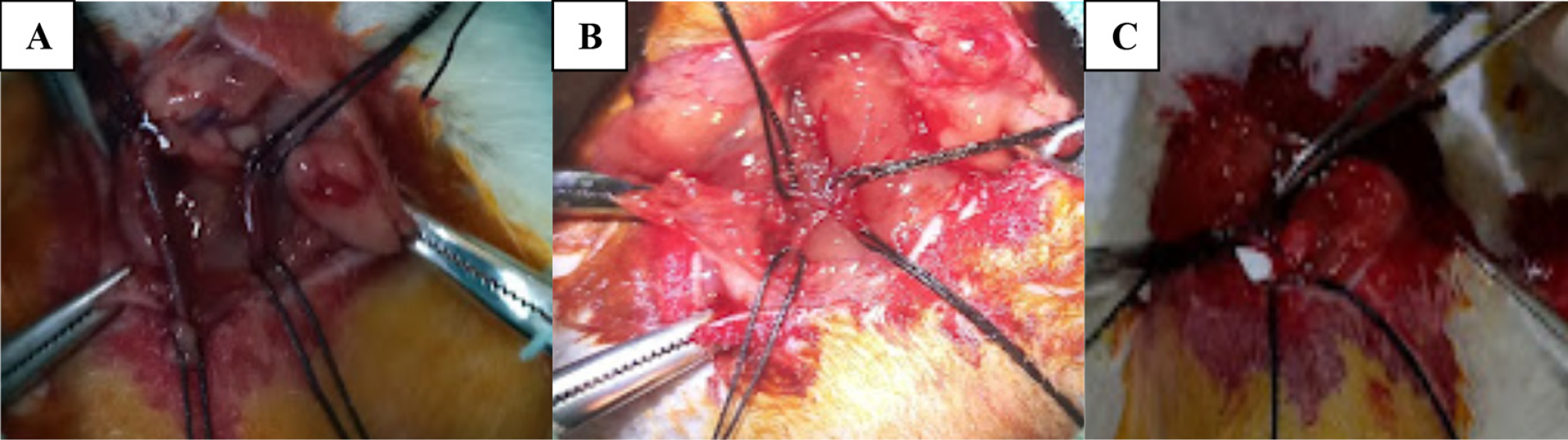
AVF: Arteriovenous fistula
On day 28 of the experiment, all rats were sacrificed by cervical dislocation under general anesthesia. Anastomotic lines were immediately collected for histopathological analysis and kept in 10% buffered formaldehyde until analysis.
Statistical Analysis
Histopathological Analysis
After euthanasia, AVF region samples obtained under general anesthesia were stored in paraformaldehyde for 48 hours. Then, they were washed under water overnight and exposed to increasing alcohol series. Finally, the samples were washed 3 times with toluene for transparency and embedded in paraffin (Slee, MPS, Mainz, Germany). Four µm sections were taken from paraffin blocks and stained with hematoxylin-eosin according to the kit protocol. Stained slides were evaluated with an Olympus BX40 (Olympus, Japan) microscope for vascular proliferation, fibroblastic cell proliferation and fibrosis parameters (0=none, 1=low, 2=moderate, 3=high) by a blinded investigator. Vascular proliferation and fibroblastic cell scores were considered together as maturation scores and were included in statistical evaluation.
The obtained data were analyzed within the scope of SPSS 24.0 package program. Differences between groups were determined by Kruskal-Wallis, and comparisons between two groups were determined by post-hoc Tukey HSD tests. Statistical significance was accepted as p<0.05. Data were summarized with mean ± standard deviation (mean ± SD).
RESULTS
Our study findings include material characterization and tissue analysis results performed on the AVF anastomosis line obtained from rats in vivo. There was no loss of rats during the entire study.
In vitro and In vivo Biodegradation Findings
In vitro biodegradation was approximately 85-90% complete on day 28 of the assay. In vivo macroscopic observation showed that the implanted biomaterial was not completely degraded in 2 animals from the PLGA group.
In vitro Drug Release Profile Findings
According to in vitro drug release assay, PLGA and MT were released at almost the same timepoint at 220 nm wavelength.
Hematoxylin-Eosin Staining Findings
Vascular proliferation and fibroblastic cell proliferation parameters were evaluated as the level of maturation with hematoxylin-eosin staining results. According to histopathological scoring, a significant increase was determined in the MT/PLGA (3.67±0.25) group compared to the Sham group (p<0.05). On the other hand, there was no significant difference between the MT/PLGA group and the PLGA (3.16±0.65) group (p>0.05). However, no significant difference was found between the Sham (2.4±0.4) and PLGA groups (, ). The data of these examinations are given in .
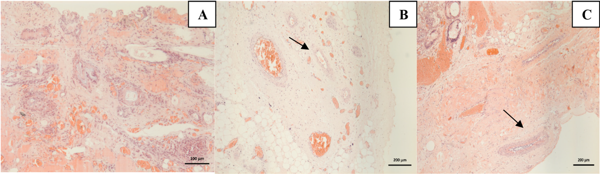
MT/PLGA: Melatonin/Polylactic-co-glycolic acid
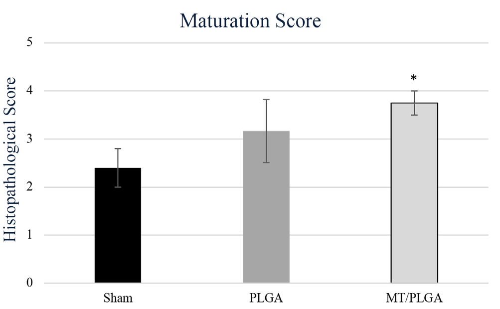
MT/PLGA: Melatonin/Polylactic-co-glycolic acid
Table 1. Maturation score and fibrosis assessment according to groups
Sham | PLGA | MT/PLGA | ||||||
|---|---|---|---|---|---|---|---|---|
Rat number | Maturation score | Fibrosis | Rat number | Maturation score | Fibrosis | Rat number | Maturation score | Fibrosis |
1 | 1 | 0 | 1 | 3 | 0 | 1 | 4 | 1 |
2 | 2 | 0 | 2 | 3 | 0 | 2 | 3 | 1 |
3 | 2 | 1 | 3 | 1 | 2 | 3 | 4 | 1 |
4 | 0 | 1 | 4 | 2 | 0 | 4 | 4 | 1 |
5 | 2 | 1 | 5 | 1 | 0 | 5 | 3 | 2 |
6 | 1 | 0 | 6 | 3 | 2 | 6 | 4 | 1 |
MT/PLGA: Melatonin/Polylactic-co-glycolic acid
Another parameter examined histopathologically was fibrosis. According to the results obtained, no significant difference was detected in terms of fibrosis between the MT/PLGA (1.25±0.25), PLGA (0.83±0.40) and Sham (0.2±0.2) groups (p>0.05).
DISCUSSION
Functional vascular access is the lifeline for patients with terminal CKD. For this purpose, AVF anastomoses are commonly used by creating an arterial and venous junction. However, the tendency for AVFs to fail before and after hemodialysis makes it impossible to provide permanent vascular access. Lack of vascular maturation significantly increases the risk of morbidity in patients with terminal kidney disease. Therefore, in addition to new surgical methods to provide long-term usable and functional vascular access, the urgent need for supportive applications for rapid maturation and its permanence has increased. For this purpose, our study aimed to develop a new method for successful and permanent vascular access by examining the effectiveness of MT-loaded PLGA nanofibers on AVF maturation.
In controlled release studies, it is possible to optimize the biomaterial produced in accordance with the healing process of the pathological mechanism. For this purpose, various synthetic or naturally sourced polymeric materials are used, . In recent years, there are biomaterials developed in the field of tissue engineering and biomaterials that have successfully completed experimental processes for use in the treatment of many diseases. Among these, synthetic biomaterials have become more preferred due to their lack of immunogenicity. Among these, PLGA is more preferred due to its biocompatibility, biodegradability and ability to mimic the extracellular matrix, which is higher than other natural and synthetic polymers. PLGA is a polymeric material approved by the FDA and is a type of synthetic polymer widely used in the field of tissue engineering due to its biocompatibility, biodegradability and extracellular matrix mimicry properties. PLGA provides antiadhesive effects and is a good choice for loading several different molecules and agents. Biodegradation of PLGA nanofibers can be optimized according to the needs of specific tissue healing-. In our study, we aimed to develop PLGA nanofibers with a structure in the form of a covering membrane. For this purpose, a biodegradable barrier containing MT and providing controlled release of MT for AVF maturation and vascular proliferation was produced, and its in vitro release and degradation profile and in vivo efficacy were determined. For this purpose, firstly PLGA nanofibers were produced and a matrix containing 1 mg MT per 1x3 cm was developed. In vitro experiments were conducted on this matrix to investigate MT release and biodegradation of its mass. As a result of In vitro studies, it was revealed that nanofibers lost 85-90% of their mass on the 28th day.
Maturation of AVF anastomoses involves complex steps including vascular repair, neointimal hyperplasia and inflammation. In these processes, the success of AVF depends on vascular proliferation and rapid fibroblastic cell proliferation. When vascular proliferation and fibroblastic cell proliferation processes are suppressed, abnormal vascular remodeling is observed. This leads to primary vascular access failure. Therefore, it is critical to ensure vascular proliferation and fibroblastic cell proliferation in the AVF anastomosis line. In the in vivo part of our study, histopathological examinations were performed with hematoxylin-eosin staining on tissue samples taken from rats. Hematoxylin-eosin staining results and vascular proliferation and fibroblastic cell proliferation parameters were evaluated in terms of maturation level. According to the histopathological scoring, a significant increase was determined in the MT/PLGA group compared to the Sham group (p<0.05). It is possible to say that MT, which was released from MT-loaded PLGA nanofibers for 28 days, provides vascular proliferation and supports AVF maturation by increasing fibroblastic cell proliferation. Studies have shown that MT regulates vascular proliferation by suppressing abnormal proliferation of smooth muscle cells, . The histopathological findings of our study suggest that MT increases vascular proliferation to provide AVF maturation and prevents intimal hyperplasia. It is likely that MT achieves this effect by increasing nitric oxide release on vascular smooth muscle and reducing oxidative stress. However, a signaling pathway or molecular mechanism in this direction was not examined in our study.
Among tissue engineering studies, considering the current literature, controlled release agents have been used to suppress intimal hyperplasia and increase vascular proliferation in biomaterial studies to ensure maturation of AVF anastomoses. In one of these studies, the vascular maturation effects of vascular endothelial growth factor (VEGF) and platelet-derived growth factor (PDGF) loaded PLGA nanofibers were evaluated. The results of this study showed that VEGF and PDGF loaded PLGA nanofibers increased vascular maturity in the first 21 days of the experiment. In another study, angiogenic factor-loaded PLGA nanofibers were implanted subcutaneously in mice to study subcutaneous vascular formation. Application of nitric oxide-releasing nanomatrix gel to rats with AVFs provided better results in outward remodeling and inhibited intimal hyperplasia at 28 days. Rapamycin-loaded nanofibrous membranes were used to evaluate the maturation effects of the material in an animal model of AVF anastomosis. After 4 weeks, the anastomosis sites were harvested and evaluated for further analysis. As a result of this study, it was stated that rapamycin-loaded nanofibrous membranes reduced intimal hyperplasia and facilitated AVF maturation. The findings obtained from our study suggest that MT-loaded PLGA nanofibers increased vascular proliferation on day 28 and provided AVF maturation. The sustained release of MT in the AVF line with a biocompatible biomaterial for 28 days indicates that it contributes to vascular proliferation in addition to its anti-inflammatory and antioxidant effects.
In our study, another parameter investigated to investigate the effects of MT-loaded PLGA nanofibers on AVF maturation was fibrosis. Fibrosis is the accumulation of extracellular collagen matrix in the vascular intima. The extent to which the presence of medial fibrosis indicates the success of the anastomosis for AVF fistula maturation is still a matter of debate in the literature. There are studies showing that increased arterial fibrosis and venous fibrosis ensure the success of AVF maturation, and there are study results showing that increased fibrosis causes AVF immaturity, . In our study, no significant results were found between the experimental groups in terms of fibrosis evaluations made from vascular sections.
In our study, we used MT-loaded PLGA nanofibers as a cover material around the AVF line. Our results revealed that MT increased vascular maturity in the early stages of remodeling. In addition, MT’s excellent antioxidant andante-inflammatory properties suggest that it contributes to maturation by preventing oxidative and inflammatory damages at the surgical site and anastomosis line. PLGA seems to be a good option for loading different agents in vascular tissue engineering.
In summary, it was observed that the MT-loaded PLGA biomaterial developed in our project initiated AVF maturation on the 28th day by providing vascular proliferation at both macroscopic and microscopic levels.
Study Limitations
In our study, biomaterial production was optimized and the effectiveness of the produced biomaterial on AVF maturation was investigated with in vivo experiments. In our study, histopathological analysis was performed only on the tissue obtained from the anastomosis line.
CONCLUSION
However, it has not been determined by which molecular signaling mechanism MT exerts these effects. In addition, biodegradation after degradation of MT-loaded PLGA nanofibers has only been determined macroscopically. It is aimed to eliminate these limitations with further research and experimental planning.
Footnotes
Ethics
Ethics Committee Approval: Ethical approval for this study was obtained from the Local Ethics Committee for Animal Experiments at Çanakkale Onsekiz Mart University (decision number: 2022/01-03, date: 21.01.2022).
Informed Consent: All procedures were carried out in accordance with the Guide for the Care and Use of Laboratory Animals.
Authorship Contributions
Surgical and Medical Practices: D.A., S.Ş, C.A., S.A., M.U.J., Concept: A.O., S.S., Design: A.O., D.A., Data Collection or Processing: S.S., D.A., C.A., Analysis or Interpretation: Ö.Y., C.A., Literature Search: D.A., Writing: D.A.
Conflict of Interest: No conflict of interest was declared by the authors.
Financial Disclosure: This study was supported by Çanakkale Onsekiz Mart University Experimental Research Coordination Unit with the THD-2022-3967 project number.
References
Turner JM, Bauer C, Abramowitz MK, Melamed ML, Hostetter TH. Treatment of chronic kidney disease. Kidney Int. 2012;81:351-62.
Carminatti M, Tedesco-Silva H, Silva Fernandes NM, Sanders-Pinheiro H. Chronic kidney disease progression in kidney transplant recipients: a focus on traditional risk factors. Nephrology (Carlton). 2019;24:141-7.
Jadoul M, Aoun M, Masimango Imani M. The major global burden of chronic kidney disease. Lancet Glob Health. 2024;12:e342-e343.
Manov JJ, Mohan PP, Vazquez-Padron R. Arteriovenous fistulas for hemodialysis: brief review and current problems. J Vasc Access. 2022;23:839-846.
Fila B, Ibeas J, Tey RR, Lovčić V, Zibar L. Arteriovenous fistula for haemodialysis: the role of surgical experience and vascular access education. Nefrología. 2016;3689-94.
Farrington CA, Cutter G, Allon M. Arteriovenous fistula nonmaturation: what’s the immune system got to do with it? Kidney360. 2021;2:1743.
Sabiu G, Gallieni M. Pathophysiology of arteriovenous fistula maturation and nonmaturation. Clin J Am Soc Nephrol. 2022;18:8.
Gameiro J, Ibeas J. Factors affecting arteriovenous fistula dysfunction: a narrative review. J Vasc Access. 2020;21:134-47.
Thomas AC, Campbell GR, Campbell JH. Advances in vascular tissue engineering. Cardiovascular Pathology. 2003;12:271-6.
Han J, Lazarovici P, Pomerantz C, Chen X, Wei Y, Lelkes PI. Co-electrospun blends of PLGA, gelatin, and elastin as potential nonthrombogenic scaffolds for vascular tissue engineering. Biomacromolecules. 2011;12:399-408.
Jeong SI, Kim SY, Cho SK, Chong MS, Kim KS, Kim H, et al. Tissue-engineered vascular grafts composed of marine collagen and PLGA fibers using pulsatile perfusion bioreactors. Biomaterials. 2007;28:1115-22.
Gökçe ON, Aykora D, Danışman M, Demircan N, Ermiş H, Gürbüz M. Development of melatonin-embedded PLGA-PEG6000 nanofiber biomaterial, and investigation of the effects on abdominal adhesion formation. 2023;39:63-75.
Lv N, Hou M, Deng L, Hua X, Zhou X, Liu H, et al. A sponge-like nanofiber melatonin-loaded scaffold accelerates vascularized bone regeneration via improving mitochondrial energy metabolism. Mater Today Bio. 2024;26:101078.
Wang S, Li J, He Y, Ran Y, Lu B, Gao J, et al. Protective effect of melatonin entrapped PLGA nanoparticles on radiation-induced lung injury through the miR-21/TGF-β1/Smad3 pathway. Front Bioeng Biotechnol. 2021;602:120584.
Mirza-Aghazadeh-Attari M, Mihanfar A, Yousefi B, Majidinia M. Nanotechnology-based advances in the efficient delivery of melatonin. Cancer Cell Int. 2022;22:1-11.
Guzmán-Soria A, Moreno-Serna V, Canales DA, García-Herrera C, Zapata PA, Orihuela PA. Effect of Electrospun PLGA/Collagen scaffolds on cell adhesion, viability, and collagen release: potential applications in tissue engineering. Polymers. 2023;15:1079.
Lee S, Kim HJ, Cho HB, Kim HR, Lee S, Park JI, et al. Melatonin loaded PLGA nanoparticles effectively ameliorate the in vitro maturation of deteriorated oocytes and the cryoprotective abilities during vitrification process. Biomater Sci. 2023;11:2912-23.
Jarrar H, Çetin Altındal D, Gümüşderelioğlu M. Effect of melatonin/BMP-2 co-delivery scaffolds on the osteoclast activity. J Mater Sci Mater Med. 2021;32.
Mendes L, Queiroz M, Sena CM. Melatonin and vascular function. Antioxidants. 2024;13:747.
Ait Abdellah S, Gal C, Guinobert I, Bardot V, Raverot V, Vitacca A, et al. Melatonin bioavailability after oral administration of a new delayed-release form in healthy male volunteers. Drugs R D. 2024;24:415-23.
Gökçe ON, Aykora D, Danışman M, Demir U, Aydeğer C, Suner SC, et al. Development of melatonin-embedded PLGA-PEG6000 nanofiber biomaterial, and investigation of the effects on abdominal adhesion formation. J Bioact Compat Polym. 2023;39:63-75.
Wang S, Li J, He Y, Ran Y, Lu B, Gao J, et al. Protective effect of melatonin entrapped PLGA nanoparticles on radiation-induced lung injury through the miR-21/TGF-β1/Smad3 pathway. Int J Pharm. 2021;602:120584.
Farid A, Michael V, Safwat G. Melatonin loaded poly(lactic-co-glycolic acid) (PLGA) nanoparticles reduce inflammation, inhibit apoptosis and protect rat’s liver from the hazardous effects of CCL4. Sci Rep. 2023;13:16424.
Das SK, Chakraborty S, Naskar S, Rajabalaya R. Techniques and methods used for the fabrication of bionanocomposites. Bionanocomposites in Tissue Engineering and Regenerative Medicine. Published online. 2021:17-43.
Ramchandani M, Pankaskie M, Robinson D. The influence of manufacturing procedure on the degradation of poly(lactide-co-glycolide) 85:15 and 50:50 implants. Journal of Controlled Release. 1997;43:161-73.
Jusu SM, Obayemi JD, Salifu AA, Nwazojie CC, Uzonwanne V, Odusanya OS, et al. Drug-encapsulated blend of PLGA-PEG microspheres: in vitro and in vivo study of the effects of localized/targeted drug delivery on the treatment of triple-negative breast cancer. Sci Rep. 2020;10:1-23.
Zheng C, Zhou Y, Huang C, Zhang Z, Liu Y, Xu Y. Establishment of a rat autogenous arteriovenous fistula model following 5/6 nephrectomy. Exp Ther Med. 2015;10:219-24.
Alpers CE, Imrey PB, Hudkins KL, Wietecha TA, Radeva M, Allon M, et al. Histopathology of veins obtained at hemodialysis arteriovenous fistula creation surgery. J Am Soc Nephrol. 2017;28:3076-88.
Applewhite B, Andreopoulos F, Vazquez-Padron RI. Periadventitial biomaterials to improve arteriovenous fistula and graft outcomes. J Vasc Access. 2024;25:713-27.
Vazquez-Padron RI, Duque JC, Tabbara M, Salman LH, Martinez L. Intimal hyperplasia and arteriovenous fistula failure: looking beyond size differences. Kidney360. 2021;2:1360.
Lee EJ, Kasper FK, Mikos AG. Biomaterials for tissue engineering. Ann Biomed Eng. 2013;42:323.
Hines DJ, Kaplan DL. Poly(lactic-co-glycolic) acid-controlled-release systems: experimental and modeling insights. Crit Rev Ther Drug Carrier Syst. 2013;30:257-76.
Liu S, Yu JM, Gan YC, Qiu XZ, Gao ZC, Wang H, et al. Biomimetic natural biomaterials for tissue engineering and regenerative medicine: new biosynthesis methods, recent advances, and emerging applications. Military Med Res. 2023;10:1-30.
Gentile P, Chiono V, Carmagnola I, Hatton P V. An overview of poly(lactic-co-glycolic) acid (PLGA)-based biomaterials for bone tissue engineering. Int J Mol Sci. 2014;15:3640-59.
Sadat Tabatabaei Mirakabad F, Nejati-Koshki K, Akbarzadeh A, Yamchi MR, Milani M, Zarghami N, et al. PLGA-based nanoparticles as cancer drug delivery systems. Asian Pac J Cancer Prev. 2014;15:517-35.
Wang B, Lu G, Song K, Chen A, Xing H, Wu J, et al. PLGA-based electrospun nanofibers loaded with dual bioactive agent loaded scaffold as a potential wound dressing material. Colloids Surf B Biointerfaces. 2023;231:113570.
Lee JY, Bashur CA, Goldstein AS, Schmidt CE. Polypyrrole-coated electrospun PLGA nanofibers for neural tissue applications. Biomaterials. 2009;30:4325.
Kim MJ, Kim JH, Yi G, Lim SH, Hong YS, Chung DJ. In vitro and in vivo application of PLGA nanofiber for artificial blood vessel. Macromol Res. 2008;16:345-52.
Siddiqui MA, Ashraff S, Santos D, Carline T. An overview of AVF maturation and endothelial dysfunction in an advanced renal failure. Ren Replace Ther. 2017;3:1-6.
Hyun BK, Kim MK, Park KG. Melatonin inhibits vascular smooth muscle cell proliferation and apoptosis through upregulation of Sestrin2. Endocrine Abstracts. 2020;70.
Lv D, Cui PL, Yao SW, Xu YQ, Yang ZX. Melatonin inhibits the expression of vascular endothelial growth factor in pancreatic cancer cells. Chin J Cancer Res. 2012;24:310.
Mendes L, Queiroz M, Sena CM. Melatonin and vascular function. Antioxidants. 2024;13:747.
Del Vento F, Poels J, Vermeulen M, Ucakar B, Giudice MG, Kanbar M, et al. Accelerated and ımproved vascular maturity after transplantation of testicular tissue in hydrogels supplemented with VEGF- and PDGF-loaded nanoparticles. Int J Mol Sci. 2021;22:5779.
Somarathna M, Hwang PT, Millican RC, Alexander GC, Isayeva-Waldrop T, Sherwood JA, et al. Nitric oxide releasing nanomatrix gel treatment inhibits venous intimal hyperplasia and improves vascular remodeling in a rodent arteriovenous fistula. Biomaterials. 2022;280:121254.
Rong D, Wang T, Liu X, Pan X. The study of rapamycin nanofibrous membrane for preventing arteriovenous fistula stenosis. Mater Des. 2024;245:113297.
Shiu YT, Litovsky SH, Cheung AK, Pike DB, Tey JCS, Zhang Y, et al. Preoperative vascular medial fibrosis and arteriovenous fistula development. Clin J Am Soc Nephrol. 2016;11:1615-23.
Martinez L, Duque JC, Tabbara M, Paez A, Selman G, Hernandez DR, et al. Fibrotic venous remodeling and nonmaturation of arteriovenous fistulas. J Am Soc Nephrol. 2018;29:1030-40.
Allon M, Litovsky S, Young CJ, Deierhoi MH, Goodman J, Hanaway M et al. Medial fibrosis, vascular calcification, ıntimal hyperplasia, and arteriovenous fistula maturation. Am J Kidney Dis. 2011;58:437-43.