
Abstract
Aim
High glucose and inflammation facilitate oral squamous cell carcinoma (OSCC) progression. The glucose transporter solute carrier family 2 member 3 (SLC2A3) and the nucleotide-binding oligomerization domain, leucine-rich repeat-containing family pyrin domain containing 3 (NLRP3), contribute to chronic inflammation and poor prognosis. This study investigates the regulatory effects of the antidiabetic drug empagliflozin (emp) on SLC2A3 and NLRP3 in OSCC and their impact on cancer cell growth.
Materials and Methods
Human OSCC cell lines, SCCL-MT1 and UPSI-SCC-131, were cultured in Dulbecco’s Modified Eagle Medium with supplements. Emp, lipopolysaccharide, and adenosine triphosphate were used for treatments. Real-time polymerase chain reaction quantified SLC2A3 and NLRP3 expressions. Enzyme-linked immunosorbent assay measured interleukin-1beta (IL-1b) levels, and cell proliferation was assessed using the xCELLigence system. Migration was evaluated via a scratch wound assay. Protein-protein interactions were predicted using the STRING database. Data analysis was conducted using GraphPad Prism.
Results
Emp, an SGLT2 inhibitor, significantly reduced SLC2A3 (p<0.0001, p=0.0002; respectively) and NLRP3 expression (p=0.0008; p=0.0006; respectively), leading to decreased IL-1b release (p=0.0190, p<0.0001; respectively), proliferation (p=0.024; p<0.0001; respectively), and migration (p=0.0021; p=0.0004, respectively) in SCCL-MT1 and UPSI-SCC131 OSCC cell lines. These findings suggest emp’s potential as a therapeutic agent for OSCC by targeting glucose metabolism and inflammation.
Conclusion
These findings suggest that emp effectively modulates glucose metabolism and inflammation in OSCC by inhibiting SLC2A3 and NLRP3 expression and IL-1b release, thereby reducing cancer cell proliferation and migration, highlighting its possible role as a treatment option for managing OSCC progression.
Keywords: Oral squamous cell carcinoma, SLC2A3, NLRP3, empagliflozin
INTRODUCTION
Elevated glucose metabolism and inflammatory processes are two key factors that facilitate tumor growth, metastasis, and resistance to therapy in oral squamous cell carcinoma (OSCC). Cancer cells must adapt to rapid microenvironmental changes, such as hypoxia, nutrient scarcity, and acidic pH, particularly when oxidative phosphorylation alone cannot sustain energy production. To compensate, they reprogram glucose metabolism to ensure sufficient adenosine triphosphate (ATP) production and meet biosynthetic demands essential for cell proliferation, . This metabolic shift enables cancer cells to generate ATP and biosynthetic precursors via aerobic glycolysis, leading to significantly increased glucose uptake in highly proliferative tumor cells, . This adaptation supplies the substrates necessary for cancer cell proliferation and contributes to tumor progression, metastasis, and long-term survival, .
Additionally, elevated glucose levels in cancer cells can induce reactive oxygen species, which consequently drive the formation of cytoplasmic multiprotein complexes such as the nucleotide-binding oligomerization domain, leucine-rich repeat-containing family pyrin domain containing 3 (NLRP3). This protein complex, known as the inflammasome, activates inflammatory responses by promoting cytokine secretion, such as pro-inflammatory interleukin-1beta (IL-1b), and releasing it into the extracellular space, thereby intensifying cellular inflammation.
Metabolic reprogramming of cancer cells is primarily regulated by hypoxia-inducible factor-1a , which controls the expression of metabolic enzymes, including glucose transporters (GLUTs), . Given this metabolic reliance, targeting glucose metabolism offers a promising approach to enhance therapeutic efficacy, . Among the GLUT family, GLUT3 plays a crucial role in maintaining basal glucose transport, particularly in cells requiring high glucose affinity for survival. In OSCC tumors, GLUT3 expression, encoded by the solute carrier family 2 member 3 (SLC2A3) gene, has been linked to poor prognosis and an aggressive cancer phenotype due to enhanced glucose metabolism, . Moreover, increased NLRP3 expression in OSCC has been associated with tumor progression and metastasis.
Given these findings, antidiabetic drugs have been investigated for their potential to modulate cancer cell metabolism and inflammatory responses. While studies have highlighted the anticancer effects of anti-diabetic drugs, including metformin, the role of sodium-glucose cotransporter-2 inhibitors, such as empagliflozin (emp), in cancer treatment remains poorly understood-. Although emp has been reported to suppress NLRP3 in obesity models, its impact on NLRP3 expression remains largely unknown in cancer. Thus, this study examined the influence of emp on SLC2A3, which encodes GLUT3, and NLRP3 in OSCC cell lines. Additionally, we explored the role of Emp-mediated SLC2A3-NLRP3 modulation in OSCC cell proliferation.
MATERIALS AND METHODS
Cells and Reagents
The human recurrent oral cavity squamous cell carcinoma cell line, SCCL-MT1, was generously provided by Bursa University, while the UPSI-SCC-131 OSCC cell line was a gift from (Bursa University). Cells were cultured in Dulbecco’s Modified Eagle Medium-high glucose medium supplemented with fetal bovine serum (10%), antibiotic solution (penicillin & streptomycin, 50 U/mL), L-glutamine (2 mM), and sodium pyruvate (1 mM) and maintained under standard culture conditions (37 °C, 5% CO2, humidified atmosphere). All experiments were conducted using passage 15 cells.
The cell culture media was from HyClone (Logan, UT, USA), while the supplements were provided by BIOCHROME (Berlin, Germany). Lipopolysaccharide (LPS) from Escherichia coli O111:B4 was purchased from Sigma (St. Louis, MO, USA), ATP was purchased from InvivoGen (San Diego, CA, USA) emp was supplied by Boehringer Ingelheim (Biberach, Germany).
Real-Time Polymerase Chain Reaction
Total RNA was isolated by the Quick-RNA Miniprep Kit (Zymo Research, Orange, CA, USA). The concentration and purity of RNA were evaluated by analyzing the A260/280 and A260/230 ratios with a Beckman Coulter DU 730 spectrophotometer (Brea, CA, USA). The ProtoScript M-MuLV First Strand cDNA Synthesis Kit (New England Biolabs, Ipswich, MA, USA) was used for reverse transcription. The expression level of SLC2A3 was analyzed using the TaqMan™ Gene Expression Assay (Thermo Fisher Scientific, USA), while NLRP3 expression was analyzed with primers reported by Tezcan et al.. Actin-beta (ACTB) served as the reference gene for normalization. For SLC2A3, the quantitative polymerase chain reaction (qPCR) reaction was performed according to the manufacturer’s instructions using TaqMan® Gene Expression Assays (Applied Biosystems, Foster City, CA, USA). For NLRP3 and ACTB, qPCR was carried out in a 10 µL reaction volume containing 200 ng of cDNA, 10 µM of each primer, and Luna® Universal qPCR Master Mix (New England Biolabs, Ipswich, MA, USA). qPCR was performed on a StepOnePlus™ real time-PCR system (Thermo Fisher Scientific, Waltham, MA, USA) with the following cycling conditions: initial denaturation at 95 °C for 2 minutes, followed by 40 cycles of denaturation at 95 °C for 10 seconds and annealing/extension at 56 °C for 60 seconds. Data analysis, including Ct value determination and fold change calculations,
was conducted using the StepOnePlus™ software and the
2^−ΔCt method. For each comparison group, experiments were performed with three independent biological replicates, each analyzed in technical duplicate.
Enzyme-Linked Immunosorbent Assay (ELISA)
A sandwich ELISA approach (SunRed Bio, Shanghai, China) was employed, utilizing a biotin-labeled detection antibody specific to the target IL-1b. Samples and standards were incubated in wells pre-coated with a capture antibody for 1 hour at 37 °C, followed by three washes with phosphate-buffered saline (PBS). The biotin-labeled detection antibody was added and incubated for 30 minutes, followed by the streptavidin-horseradish peroxidase conjugate. After a 30-minute incubation with the substrate solution in the dark, color development was measured at 450 nm (reference: 650 nm) using a microplate reader. Each comparison group was analyzed in three biological repeats, each analyzed in technical duplicates. The results were calculated based on the standard curve generated from IL-1b reference standards.
Dynamic Cell Proliferation Analysis
SCCL-MT1 and UPSI-SCC-131 cells (5 × 10 per well) were plated into an E-Plate 16 (ACEA Biosciences, San Diego, CA, USA). Cell growth was perpetually monitored every 30 minutes over a 96-hour period using the xCELLigence real-time cell analysis (RTCA) dual purpose system (ACEA Biosciences, San Diego, CA, USA), which measures electrical impedance as an indicator of cell viability and proliferation. Background impedance was recorded before cell seeding to ensure accurate normalization. For each of the two different cell lines, the experiment was conducted with three independent biological replicates per comparison group. The resulting proliferation curves were generated and analyzed using the RTCA Software (ACEA Biosciences).
In-vitro Wound Healing Scratch Assay
A scratched wound-healing assay was conducted to visualize cell migration. Once the monolayer achieved confluence, a scratch was introduced using a 1000 μL pipette tip, creating a standardized wound area. Detached cells and debris were carefully removed by washing with PBS. Images of the wounded region were recorded after scratching and again 24 hours post-treatment using an inverted microscope (Nikon, Tokyo, Japan). Wound closure was quantified by measuring the wound area at both time points using ImageJ v1.54f software (National Institutes of Health, Bethesda, MD, USA). The experiment was performed in three biological replicates per comparison group, with each replicate analyzed in technical duplicate to ensure reproducibility.
Statistical Analysis
Protein-protein interactions were evaluated through the STRING v12.0, (https://string-db.org/) (accessed on February 24, 2025). Statistical analysis was achieved by GraphPad Prism (v14.4.0) (GraphPad Software, Boston, MA, USA). Data were confirmed to follow a normal distribution based on Q-Q plot analysis and Shapiro-Wilk tests, with p>0.05 indicating no significant deviation from normality. Homogeneity of variances was evaluated using Levene’s test. Group differences were analyzed using one-way ANOVA, followed by Tukey’s post-hoc test for pairwise comparisons to control for Type I error inflation due to multiple testing. Results are reported as mean ± standard error, with significance defined as p<0.05. All statistical tests were conducted with at least three independent biological replicates to ensure data robustness and reproducibility.
RESULTS
Empagliflozin Suppressed SLC2A3 Expression in OSCC Cells
To establish an inflammation-induced OSCC cell model, SCCL-MT1 and UPSI-SCC131 cells were exposed to 1 µg/mL LPS for 3 hours. Additionally, cells were treated with 500 nM Emp, an SGLT2 inhibitor, for 24 hours to suppress glucose transport as described in our previous study. Our results demonstrated that emp significantly downregulated SLC2A3 RNA expression in both SCCL-MT1 (p<0.0001), and UPSI-SCC131 cells (p=0.0002). Moreover, SLC2A3 expression was further reduced in LPS-induced cells when subsequently exposed to Emp, compared to cells treated with LPS alone (p<0.0001) ( A, B).
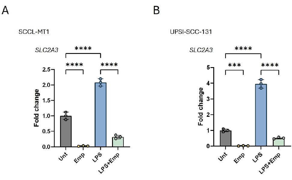
Unt: Untreated, LPS: Lipopolysaccharide, emp: Empagliflozin, OSCC: Oral squamous cell carcinoma
Empagliflozin Reduced NLRP3 Expression and Suppresses IL-1β Release in LPS-Induced Inflammation
Genetic interaction analysis predicted that both SLC2A3 and NLRP3 interact with IL-1b (). Supporting these findings, emp reduced NLRP3 expression in SCCL-MT1 (p=0.0008) and UPSI-SCC131 cells (p=0.0006) (, ). Similarly, in LPS-treated SCCL-MT1 and UPSI-SCC131 cells, the emp treatment led to a decrease in NLRP3 expression than the cells triggered by LPS-only (p<0.0001). To evaluate the effect of emp on IL-1b release upon inflammasome activation, cells were treated with 1 µg/mL LPS for 3 hours, followed by 24-hour emp treatment and 25-minute exposure to 5 mM ATP (). IL-1b release was significantly increased in inflammasome-activated cells compared to those cells under LPS-induced inflammation conditions (p<0.0001). However, emp decreased IL-1b release both in LPS-treated cells (SCCL-MT1: p=0.0190; UPSI-SCC131) (p<0.0001) and in inflammasome-activated cells with LPS-ATP treatment (p<0.0001) (, ), suggesting that Emp, through the inhibition of SLC2A3, may also reduce NLRP3-mediated inflammatory cytokine production.
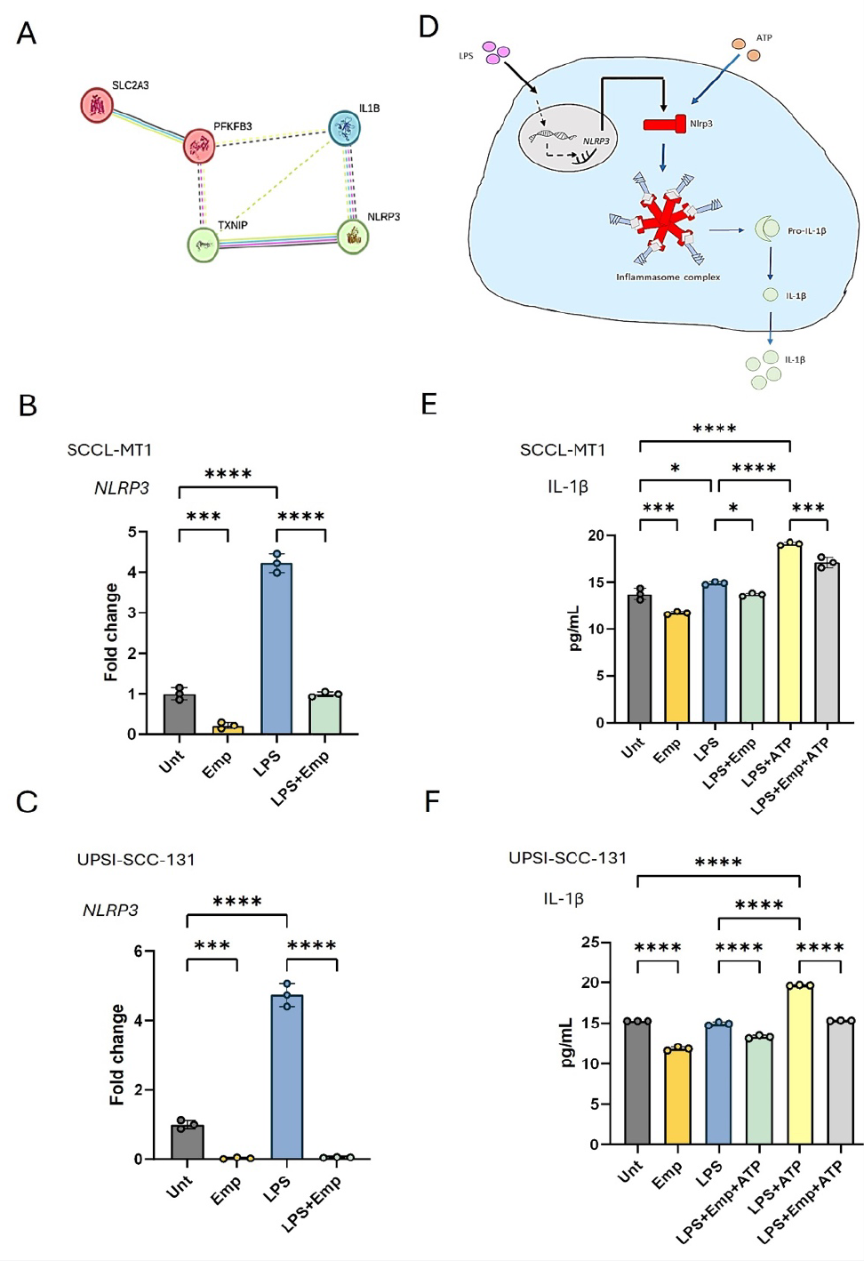
Unt: Untreated, LPS: Lipopolysaccharide, ATP: Adenosine triphosphate, emp: Empagliflozin, OSCC: Oral squamous cell carcinoma, IL-1b: Interleukin-1beta, RT-qPCR: Reverse transcription quantitative polymerase chain reaction
Empagliflozin Slowed Cell Proliferation and Migration in OSCC Cells
Emp treatment decreased the proliferation of both SCCL-MT1 and UPSI-SCC131 cell lines (, ). In contrast, LPS treatment increased cell proliferation compared to untreated controls. However, emp treatment also inhibited proliferation in LPS-pretreated cells relative to those treated with LPS alone (, ).
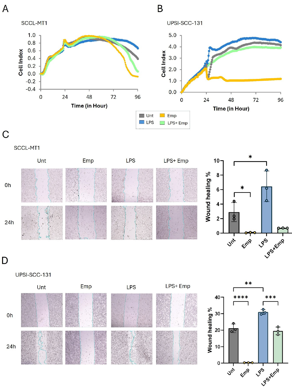
Unt: Untreated, LPS: Lipopolysaccharide, Emp: Empagliflozin, OSCC: Oral squamous cell carcinoma
Wound healing assays further supported these findings, showing that emp decreased cell migration in both SCCL-MT1 (p=0.024) and UPSI-SCC131 cells (p<0.0001) (, ). In contrast; LPS enhanced migration, accelerating cell movement (, ). Notably, emp also reduced wound healing in LPS-pretreated cells (SCCL-MT1: p=0.0021; UPSI-SCC131: p=0.0004) (, ). These results suggest that Emp-mediated suppression of SLC2A3 and NLRP3 reduces both the growth rate and invasive potential of OSCC cells, leading to a decrease in aggressive cancer cell behavior.
DISCUSSION
This study investigates the role of emp on NLRP3 inflammasome activation in SCCL-MT1 and UPSI-SCC131, OSCC cell lines. SCCL-MT1, derived from buccal mucosa, is an invasive and recurrent OSCC model with strong immunomodulatory activity. Previous studies have demonstrated that SCCL-MT1 promotes immunosuppression within the tumor microenvironment by stimulating myeloid-derived suppressor cells via IL-1b secretion, leading to effector T-cell suppression and an increase in regulatory T-cells, . Similarly, UPSI-SCC131, originating from a patient with a history of tobacco and alcohol use, is resistant to radiotherapy, . This resistance has been associated with elevated IL-1b secretion. Due to their clinically relevant characteristics such as invasiveness, immunomodulatory activity, and therapy resistance, the effects of emp on OSCC were investigated using the SCCL-MT1 and UPSI-SCC131 cell lines in this study.
Empagliflozin-Mediated Mechanistic Link Between SLC2A3 and NLRP3
IL-1b production and secretion in vitro require a two-step activation of the NLRP3 inflammasome. In the priming phase, pathogen-associated stimuli such as LPS activate Toll-like receptors, leading to NF-kB signaling and enhanced NLRP3 transcription. In the activation phase, ATP stimulates the P2X7 receptor, inducing potassium efflux and promoting NLRP3 inflammasome assembly. This process culminates in pro-caspase-1 activation and IL-1b secretion-. Therefore, we evaluated the effect of emp on NLRP3 activation by stimulating SCCL-MT1 and UPSI-SCC131 cells with LPS, followed by ATP exposure, and analyzing IL-1b secretion.
Building on these mechanistic insights, our findings demonstrate that treatment with Emp, apharmacological inhibitor of glucose transport, downregulates SLC2A3 (GLUT3) mRNA expression, indicating reduced glucose uptake in OSCC cells. Emp also suppresses NLRP3 expression and inhibits inflammasome activation, leading to decreased IL-1b secretion. These results suggest a functional link between SLC2A3 and NLRP3 in the regulation of glucose metabolism and inflammation in cancer cells.
A recent study has associated elevated SLC2A3 expression with the activation of epithelial-mesenchymal transition and NF-kB signaling pathways, contributing to poor prognosis in patients with head and neck squamous cell carcinoma. NF-kB plays a pivotal role in initiating inflammasome activation by upregulating the transcription of NLRP3 and pro-IL-1b during the priming phase. In our study, LPS was used to model pathogen-associated molecular patterns. Importantly, even in the absence of LPS stimulation, emp suppressed SLC2A3 and NLRP3 expression, along with IL-1b secretion. These findings suggest that SLC2A3-mediated glucose uptake alone may contribute to inflammasome activation. Indeed, previous studies have shown that elevated glucose transport triggers inflammatory signaling in macrophages, the cell type where NLRP3 activation is most pronounced.
To further explore this connection, we conducted a gene–gene interaction analysis, which suggested a potential mechanistic link between SLC2A3 and NLRP3 through the glycolysis-regulating enzyme 6-phosphofructo-2-kinase/fructose-2,6-bisphosphatase 3 (PFKFB3). Cancer cells overexpress GLUTs such as SLC2A3 to increase glucose influx, which elevates intracellular fructose-2,6-bisphosphate (F2,6BP) levels. This metabolite allosterically activates phosphofructokinase-1, the rate-limiting enzyme in glycolysis. PFKFB3 amplifies this effect by promoting F2,6BP synthesis, accelerating glycolytic flux and enhancing lactate production. Lactate is exported from the cell, bypassing mitochondrial oxidative metabolism and promoting reliance on aerobic glycolysis. This shift supports rapid proliferation and fosters a hypoxic tumor microenvironment. Emerging evidence indicates that PFKFB3 not only promotes glycolytic flux but also contributes to NLRP3 inflammasome activation. Suppression of PFKFB3 has been shown to reduce glycolysis, and in macrophages, NLRP3 can regulate glycolysis through PFKFB3. Moreover, PFKFB3-induced hypoxia has been reported to trigger NLRP3 activation, while downregulation of PFKFB3 reduced NLRP3 downstream targets, including caspase-1 and IL-1b, in an atherosclerosis model. Notably, LPS has been shown to enhance ROS generation induced by high glucose–associated hypoxia, thereby exacerbating NLRP3 activation.
Taken together, these findings support the hypothesis that the SLC2A3-PFKFB3-NLRP3 axis may represent a potential mechanistic link between altered glucose metabolism and inflammation in OSCC (). Although based on in silico predictions, this pathway suggests that by suppressing SLC2A3, emp may attenuate NLRP3 inflammasome activation, potentially through modulation of PFKFB3, thereby exerting both metabolic and anti-inflammatory effects in oral cancer.
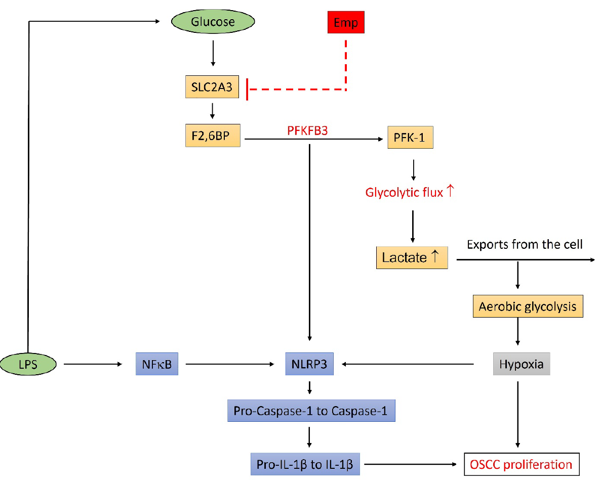
LPS: Lipopolysaccharide, emp: Empagliflozin, OSCC: Oral squamous cell carcinoma, LPS: Lipopolysaccharide, SLC2A3: Solute carrier family 2 member 3
Therapeutic Implications of Emp on OSCC
The role of the NLRP3 inflammasome in cancer remains controversial. While some studies associate NLRP3 overexpression with tumor aggressiveness and poor prognosis, others report minimal effects upon its inhibition. Our previous work using Glibenclamide and VX765, two NLRP3 inhibitors, showed only modest reductions in OSCC cell proliferation, . In contrast, emp exhibited a more pronounced anti-cancer effect, significantly decreasing OSCC cell proliferation and impairing wound healing, suggesting greater therapeutic potential through dual metabolic and inflammatory modulation.
Notably, SLC2A3 is known to promote OSCC progression through the TGF-b signaling. In addition, TGF-b signaling has been implicated in chronic inflammation through an IL-1b-driven autocrine mechanism. Although not tested in this study, emp’s reported inhibition of TGF-b signaling suggests a potential additional mechanism for its effects in OSCC, which warrants further investigation. The convergence of SLC2A3-mediated glucose transport and TGF-b–IL-1b signaling pathways may represent a novel therapeutic target axis in OSCC.
Collectively, these data support the hypothesis that glucose transport contributes to OSCC aggressiveness, and its inhibition by emp may help suppress this phenotype. Under LPS-induced inflammatory conditions, proliferation and migration rates increased but were effectively reduced by Emp. These findings highlight emp’s potential not only as an antidiabetic agent but also as a repurposed therapeutic candidate targeting metabolic-inflammation crosstalk in OSCC.
Comparison of Emp and Conventional NLRP3 Inhibitors
Mechanistically, Glibenclamide prevents inflammasome formation by blocking NLRP3 monomer assembly, while VX765 inhibits caspase-1 activation, which is required for IL-1b maturation, , . Although emp does not directly target the inflammasome complex, our results suggest it reduces NLRP3 expression by limiting glucose availability, a key upstream stimulus. Thus, emp may act earlier in the NLRP3 pathway and exert additional effects via SLC2A3-associated signaling.
Overall, this study reveals that metabolic regulation of glucose uptake plays a critical role in the progression of inflammation associated with OSCC cells. Notably, the indirect interaction between SLC2A3 and NLRP3 may contribute to shaping this inflammatory response. In this context, the suppression of proliferation and migration observed after treatment with glucose transport inhibitors such as emp likely results from the downregulation of NLRP3 expression. This reduction may limit NLRP3-associated signaling pathways involved in inflammation, thereby decreasing the secretion of pro-tumorigenic cytokines.
Indeed, the effects of emp under both basal and LPS-induced inflammatory conditions highlight the potential of glucose uptake inhibition to reduce pro-inflammatory signaling and disrupt autocrine cytokine loops in cancer cells, thereby mitigating the aggressive phenotype of OSCC.
Study Limitations
This study is an in vitro investigation conducted using two commercial cell lines. In OSCC inflammation, the inflammatory activities of non-cancerous cells within the tumor microenvironment and their interactions with cancer cells play a role. However, the experimental setup in this study consists of a homogeneous system containing only cancer cells, thereby excluding the influence of the tumor microenvironment. This represents a limitation of our study.
Furthermore, the study employed a single concentration of Emp, selected based on preliminary cytotoxicity and efficacy data. Although effective, the use of a single dose restricts dose–response interpretation and does not account for possible concentration-dependent effects or therapeutic windows. Additional experiments using multiple concentrations and time points would enhance the pharmacodynamic understanding of emp in OSCC.
Another important limitation is the lack of in vivo validation. Without animal model data, it is difficult to predict the systemic effects of Emp, its pharmacokinetics, or its potential interactions within a living organism.
Moreover, as this is not a patient-based study, the focus is solely on the effects of emp on cancer cell biology, disregarding potential variations arising from patient heterogeneity, etiological differences, and epigenetic diversity. These factors may limit the clinical generalizability and translational applicability of our findings.
CONCLUSION
In conclusion, our findings demonstrate that Emp, an SGLT2 inhibitor, suppresses SLC2A3 and NLRP3 expression, thereby reducing OSCC cell proliferation and migration. Its dual action, disrupting glucose metabolism and mitigating chronic inflammation via NLRP3 inhibition, may attenuate the aggressive phenotype of OSCC. Taken together, these insights highlight emp’s potential as a therapeutic agent for OSCC, though further studies are needed to validate its clinical efficacy.
Footnotes
Ethics
Ethics Committee Approval: The study does not involve human or animal subjects, ethical approval is not required. The relevant statement has also been included in the manuscript.
Informed Consent: This study is an in vitro study conducted using two commercially available cell lines. No biological material derived from patients or experimental animals was used, and no clinical or personal data from any patient were included.
Conflict of Interest: No conflict of interest was declared by the authors.
Financial Disclosure: The authors declared that this study received no financial support.
References
Chen Y, Han Z, Zhang L, Gao C, Wei J, Yang X, et al. TIMELESS promotes reprogramming of glucose metabolism in oral squamous cell carcinoma. J Transl Med. 2024;22:21.
Oizel K, Chauvin C, Oliver L, Gratas C, Geraldo F, Jarry U, et al. Efficient mitochondrial glutamine targeting prevails over glioblastoma metabolic plasticity. Clin Cancer Res. 2017;23:6292-6304.
Porporato PE, Dhup S, Dadhich RK, Copetti T, Sonveaux P. Anticancer targets in the glycolytic metabolism of tumors: a comprehensive review. Front Pharmacol. 2011;2:49.
Liberti MV, Locasale JW. The Warburg effect: how does it benefit cancer cells? [published correction appears in Trends Biochem Sci. 2016;41:287]. Trends Biochem Sci. 2016;41:211-218.
Levine AJ, Puzio-Kuter AM. The control of the metabolic switch in cancers by oncogenes and tumor suppressor genes [published correction appears in Science. 2012;11;336:670]. Science. 2010;330:1340-1344.
Ortega MA, De Leon-Oliva D, García-Montero C, Fraile-Martinez O, Boaru DL, de Castro AV, et al. Reframing the link between metabolism and NLRP3 inflammasome: therapeutic opportunities. Front Immunol. 2023;14:1232629.
Guo H, Callaway JB, Ting JP. Inflammasomes: mechanism of action, role in disease, and therapeutics. Nat Med. 2015;21:677-687.
Gupta S, Roy A, Dwarakanath BS. Metabolic cooperation and competition in the tumor microenvironment: implications for therapy. Front Oncol. 2017;7:68.
Semenza GL. Targeting HIF-1 for cancer therapy. Nat Rev Cancer. 2003;3:721-732.
Vander Heiden MG, DeBerardinis RJ. Understanding the intersections between metabolism and cancer biology. Cell. 2017;168:657-669.
Martinez-Outschoorn UE, Peiris-Pagés M, Pestell RG, Sotgia F, Lisanti MP. Cancer metabolism: a therapeutic perspective. Nat Rev Clin Oncol. 2017;14:113.
Yager JY. Glucose and perinatal brain injury-questions and controversies. Elsevier. 2019:141-161.
Ayala FR, Rocha RM, Carvalho KC, Carvalho AL, da Cunha IW, Lourenço SV, et al. GLUT1 and GLUT3 as potential prognostic markers for oral squamous cell carcinoma. Molecules. 2010;15:2374-2387.
Kumari A, Jha A, Tiwari A, Nath N, Kumar A, Nagini S, et al. Role and regulation of GLUT1/3 during oral cancer progression and therapy resistance. Arch Oral Biol. 2023;150:105688.
Wang H, Luo Q, Feng X, Zhang R, Li J, Chen F. NLRP3 promotes tumor growth and metastasis in human oral squamous cell carcinoma. BMC Cancer. 2018;18:500.
Dąbrowski M. Diabetes, antidiabetic medications and cancer risk in type 2 diabetes: focus on SGLT-2 Inhibitors. Int J Mol Sci. 2021;22:1680.
Dutka M, Bobiński R, Francuz T, Garczorz W, Zimmer K, Ilczak T, et al. SGLT-2 inhibitors in cancer treatment-mechanisms of action and emerging new perspectives. Cancers (Basel). 2022;14:5811.
Wu W, Wang Y, Xie J, Fan S. Empagliflozin: a potential anticancer drug. Discov Oncol. 2023;14:127.
Kocaeli AA, Tekin C, Ercelik M, Tezcan G, Aksoy SA, Kocaeli H, et al. Concurrent presence of diabetes affects the GLUT3 programming of glucose metabolism in glioblastoma. Eur Rev Med Pharmacol Sci. 2023;27:8110-8118.
Benetti E, Mastrocola R, Vitarelli G, Cutrin JC, Nigro D, Chiazza F, et al. Empagliflozin protects against diet-induced NLRP-3 inflammasome activation and lipid accumulation. J Pharmacol Exp Ther. 2016;359:45-53.
Tezcan G, Alsaadi M, Hamza S, Garanina EE, Martynova EV, Ziganshina GR, et al. Azithromycin and ceftriaxone differentially activate NLRP3 in LPS primed cancer cells. Int J Mol Sci. 2022;23:9484.
Hamza S, Garanina EE, Shkair L, Alsaadi M, Khaiboullina SF, Tezcan G. Implications of NLRP3 suppression using glibenclamide and miR-223 against colorectal cancer. Pharmaceuticals (Basel). 2024;17:299.
Russell SM, Lechner MG, Gong L, Megiel C, Liebertz DJ, Masood R, et al. USC-HN2, a new model cell line for recurrent oral cavity squamous cell carcinoma with immunosuppressive characteristics. Oral Oncol. 2011;47:810-817.
Lechner MG, Liebertz DJ, Epstein AL. Characterization of cytokine-induced myeloid-derived suppressor cells from normal human peripheral blood mononuclear cells [published correction appears in J Immunol. 2010 Nov 1;185:5668]. J Immunol. 2010;185:2273-2284.
White JS, Weissfeld JL, Ragin CC, Rossie KM, Martin CL, Shuster M, et al. The influence of clinical and demographic risk factors on the establishment of head and neck squamous cell carcinoma cell lines. Oral Oncol. 2007;43:701-712.
Sürer Şİ, Elçitepe TB, Akçay D, Daşkın E, Çalıbaşı Kocal G, Arıcan Alıcıkuş Z, et al. A promising, novel radiosensitizer nanodrug complex for oral cavity cancer: cetuximab and cisplatin-conjugated gold nanoparticles. Balkan Med J. 2021;38:278-286.
Tiwari DK, Hannen R, Unger K, Kohl S, Heß J, Lauber K, et al. IL1 pathway in HPV-Negative HNSCC cells is an indicator of radioresistance after photon and carbon ion irradiation without functional involvement. Front Oncol. 2022;12:878675.
Tengesdal IW, Dinarello CA, Marchetti C. NLRP3 and cancer: pathogenesis and therapeutic opportunities. Pharmacol Ther. 2023;251:108545.
Gurung P, Li B, Subbarao Malireddi RK, Lamkanfi M, Geiger TL, Kanneganti TD. Chronic TLR stimulation controls NLRP3 inflammasome activation through IL-10 mediated regulation of NLRP3 expression and caspase-8 activation. Sci Rep. 2015;5:14488.
Rotondo JC, Mazziotta C, Lanzillotti C, Stefani C, Badiale G, Campione G, et al. The role of purinergic P2X7 receptor in inflammation and cancer: novel molecular insights and clinical applications. Cancers (Basel). 2022;14:1116.
Zügner E, Yang HC, Kotzbeck P, Boulgaropoulos B, Sourij H, Hagvall S, et al. Differential in vitro effects of SGLT2 inhibitors on mitochondrial oxidative phosphorylation, glucose uptake and cell metabolism. Int J Mol Sci. 2022;23:7966.
Chai F, Zhang J, Fu T, Jiang P, Huang Y, Wang L, et al. Identification of SLC2A3 as a prognostic indicator correlated with the NF-kB/EMT axis and immune response in head and neck squamous cell carcinoma. Channels. 2023;17:2208928.
Liu T, Zhang L, Joo D, Sun SC. NF-kB signaling in inflammation. Signal Transduct Target Ther. 2017;2:17023.
Johnson AR, Milner JJ, Makowski L. The inflammation highway: metabolism accelerates inflammatory traffic in obesity. Immunol Rev. 2012;249:218-238.
Lebreton F, Berishvili E, Parnaud G, Rouget C, Bosco D, Berney T, et al. NLRP3 inflammasome is expressed and regulated in human islets. Cell Death Dis. 2018:25;9:726.
Lu L, Chen Y, Zhu Y. The molecular basis of targeting PFKFB3 as a therapeutic strategy against cancer. Oncotarget. 2017;24:62793-62802.
Finucane OM, Sugrue J, Rubio-Araiz A, Guillot-Sestier MV, Lynch MA. The NLRP3 inflammasome modulates glycolysis by increasing PFKFB3 in an IL-1b-dependent manner in macrophages. Sci Rep. 2019;9:4034.
Wang X, Liu X, Wu W, Liao L, Zhou M, Wang X, et al. Hypoxia activates macrophage-NLRP3 inflammasome promoting atherosclerosis via PFKFB3-driven glycolysis. FASEB J. 2024;38:e23854.
Qiu Z, He Y, Ming H, Lei S, Leng Y, Xia ZY. Lipopolysaccharide (LPS) aggravates high glucose- and hypoxia/reoxygenation-induced injury through activating ROS-dependent NLRP3 inflammasome-mediated pyroptosis in H9C2 cardiomyocytes. J Diabetes Res. 2019;17:8151836.
Moossavi M, Parsamanesh N, Bahrami A, Atkin SL, Sahebkar A. Role of the NLRP3 inflammasome in cancer. Mol Cancer. 2018;17:158.
Tezcan G, Garanina EE, Alsaadi M, Gilazieva ZE, Martinova EV, Markelova MI, et al. Therapeutic potential of pharmacological targeting NLRP3 inflammasome complex in cancer. Front Immunol. 2021;11:607881.
Jiang W, Xu S, Li P. SLC2A3 promotes tumor progression through lactic acid-promoted TGF-b signaling pathway in oral squamous cell carcinoma. PLoS One. 2024;19:e0301724.
Kang H, Seo E, Oh YS, Jun HS. TGF-b activates NLRP3 inflammasome by an autocrine production of TGF-b in LX-2 human hepatic stellate cells. Mol Cell Biochem. 2022;477:1329-1338.
Shentu Y, Li Y, Xie S, Jiang H, Sun S, Lin R, et al. Empagliflozin, a sodium glucose cotransporter-2 inhibitor, ameliorates peritoneal fibrosis via suppressing TGF-b/Smad signaling. Int Immunopharmacol. 2021;93:107374.
Zahid A, Li B, Kombe AJK, Jin T, Tao J. Pharmacological Inhibitors of the NLRP3 inflammasome. Front Immunol. 2019;10:2538.