
Abstract
Aim
In this study, it was aimed to investigate the predictive value of pan-immune-inflammation value (PIV) in postoperative saphenous vein graft patency (SVG).
Materials and Methods
Data of 300 patients with coronary artery bypass grafting (CABG) who underwent angiography between January 2022 and January 2024 were retrospectively analyzed. These patients were divided into two groups according to the presence of 50% or more stenosis in SVG (Group 1: SVG patent, Group 2: SVG not patent). We investigated PIV, neutrophil to lymphocyte ratio, platelet to lymphocyte ratio (PLR), lymphocyte to monocyte ratio, systemic inflammation response index, and systemic inflammation index. The study aimed to investigate whether there was a difference in inflammation indexes between the groups.
Results
The cut-off value of PIV >444 was associated with 72% sensitivity and 62% specificity to predict SVG disease (SVGD) in patients with CABG. Multivariate logistic regression analysis showed that PLR and PIV levels were independent predictors of SVGD, respectively [odds ratio (OR): 1.025; 95% confidence interval (CI): 1.008-1.042; p=0.003)] and (OR: 1.012; 95% CI: 1.000-1.015; p=0.004).
Conclusion
PIV and PLR may be useful predictors of SVGD, which can be easily estimated from blood tests in routine practice. As a novel inflammatory biomarker, PIV may serve as a valuable tool for risk stratification and long-term follow up in this patient population.
Keywords: Inflammation, coronary artery disease, coronary artery bypass grafting, saphenous vein graft, pan-immune-inflammation value
INTRODUCTION
Percutaneous coronary intervention (PCI) and coronary artery bypass grafting (CABG) are considered revascularization procedures, but CABG alone is the most effective treatment in some patient groups. PCI is only intended to treat flow-limiting lesions, even though non-flow-limiting stenoses cause many infarcts. PCI cannot thus be expected to reduce the number of new infarcts considerably; but, by supplying flow distal to artery occlusions, CABG may be able to accomplish so. Long-term clinical outcomes and recurring symptoms following CABG surgery are contingent upon the patency of the bypass graft and the rate of native coronary artery disease development. The most important problem after CABG is the faster progression of atherothrombotic occlusive disease in vein grafts. Inflammatory parameters are of great importance in predicting this early atherosclerosis in saphenous vein grafts (SVG). Thus, it has been discovered in several studies that choosing an arterial graft improves long-term survival and lowers the frequency of coronary angiographic procedures. In the first month following bypass surgery, thrombotic occlusion is the cause of occlusion in vein grafts; however, atherosclerosis and neointimal hyperplasia are the causes in later stages. Atherosclerosis is a chronic inflammatory vascular disease whose pathogenesis is caused by traditional and non-traditional risk factors. Genome studies have shown that innate and adaptive immune responses can promote or suppress atherosclerosis. Russell Ross originally put up the theory that atherosclerosis is an inflammatory disease in 1999 based on data showing that circulating monocytes penetrate the fatty streak as it develops. Many simple markers have been studied to predict cardiovascular mortality and stent re-stenosis, which can be obtained from biochemical parameters, .
The increase in inflammation risk markers such as neutrophil-tolymphocyte ratio (NLR) after CABG or PCI is useful in predicting cardiovascular mortality or in-stent restenosis. Many studies have demonstrated that high NLR and platelet to lymphocyte ratio (PLR) levels correlate with the severity of coronary artery disease, . Pan-immune-inflammation value (PIV) includes more comprehensive blood parameters, which makes it a better predictor in coronary artery disease than other inflamatuar indexes. In this study, it was aimed to predict saphenous vein patency with these new inflammation markers in patients with CABG.
MATERIALS AND METHODS
Data of 300 patients with CABG who underwent angiography between January 2022 and January 2024 were retrospectively analyzed (in ). A 50% or less coronary artery stenosis was considered non-critical. Patients who had previously undergone CABG were divided into two groups according to whether there was 50% or more stenosis in the SVG. Patients’ medical records were reviewed for basic demographic information, coronary angiography reports, clinical history, prescription information, and blood chemistry test findings (). The following systemic inflammation indexes were calculated from whole blood assays: [systemic inflammation response index (SIRI) (neutrophils × monocytes / lymphocytes), systemic immune inflammation index (SII) (neutrophils × platelets / lymphocytes), PIV (neutrophils × monocytes × platelets) / lymphocytes), PLR (platelets / lymphocytes ratio), lymphocyte to monocyte ratio (LMR) (lymphocytes /monocytes ratio), and NLR (neutrophils / lymphocytes ratio)]. The numbers of all blood parameters were multiplied by (×10³/µL). Patients with a history of acute coronary syndrome in the last 3 months and those with a recent history of PCI were excluded from the study. Blood samples of the patients were taken from the antecubital vein at first hospital admission after 12 hours of fasting before the angiography procedure. Patients with active infectious disease [white blood cell count (WBC)> 11 ×10³/µL or C-reactive protein (CRP)> 5mg/dL], chronic inflammatory disease (CRP> 5mg/dL or sedimentation> 20mm/hour), or clinical evidence of cancer, severe renal disease (estimated glomerular filtration rate <30 mL/min/1.73 m2), and hematological diseases were excluded from the study. Patients who underwent valve surgery together with CABG surgery were not included in the study. The study was authorized by Tekirdağ Namık Kemal University Local Ethics Committee (decision no: 2021.284.12.07, date: 28.12.2021) and was carried out by the Helsinki Declaration.
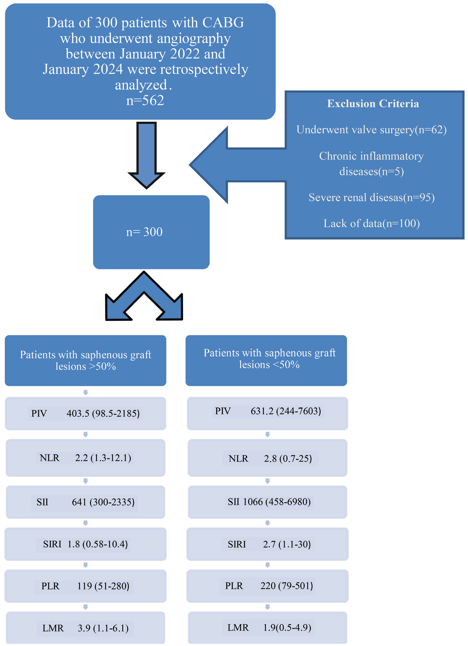
Table 1. Baseline characteristics of the groups
Variables | Group 1 (n=150) | Group 2 (n=150) | p-value |
|---|---|---|---|
Age (years) | 66.4±9.6 | 67.4±8.1 | 0.582 |
Male, n (%) | 69 (46) | 70 (46.6) | 0.986 |
Heart failure, n (%) | 10 (6.6) | 30 (20) | 0.032 |
COPD, n (%) | 12 (8) | 10 (6.6) | 0.232 |
Stroke, n (%) | 6 (4) | 4 (2.6) | 0.123 |
Hypertension, % n (%) | 50 (33.3) | 48 (32) | 0.853 |
Diabetes mellitus, n (%) | 22 (14.6) | 25 (16.6) | 0.548 |
Medical treatment | - | - | - |
Beta blocker, n (%) | 35 (23.3) | 70 (46.6) | 0.002 |
Ca-channel blocker, n (%) | 15 (10) | 20 (13.3) | 0.724 |
ACE-I/ARB, n (%) | 60 (40) | 100 (66.6) | 0.001 |
Diuretic, n (%) | 26 (17.3) | 36 (24) | 0.051 |
OAD n, (%) | 15 (10) | 18 (12) | 0.825 |
Insulin, n (%) | 20 (13.3) | 18 (12) | 0.659 |
OAD: Oral anti-diabetic drugs, ACE-I/ARB: Angiotensin-converting enzyme inhibitors/angiotensin receptor blockers, COPD: Chronic obstructive pulmonary disease, Group 1: Saphenous vein grafts patent, Group 2: Saphenous vein grafts not patent
Angiographic Analysis
After getting each patient’s informed consent, the Judkins technique was used to perform coronary angiography in normal standard projections with the required catheters. Two independent cardiologists who were blind to the patient data analyzed the coronary angiograms of 300 patients. Bypass grafts with visual stenosis of 50% or more were considered significant. These patients were divided into two groups according to the presence of 50% or more stenosis in SVG. In Group 1, there were 150 patients with less than 50% stenosis in their bypass grafts. In Group 2, there were 150 patients with significant stenosis of over 50% in their bypass grafts.
Statistical Analysis
SPSS 22.0 statistical software (SPSS Inc., Chicago, IL) was used to analyze all of the study’s data. The Kolmogorov-Smirnov test was used to evaluate data distribution. Categorical variables were reported as percentages and compared using chi-square test or Fischer’s exact test while continuous variables were expressed as mean ± standard deviation or median (minimum-maximum). Continuous data conforming to normal distribution was evaluated with the Student’s t-test, and data not compatible with normal distribution was evaluated with the Mann-Whitney U test. ROC curve analysis was used to determine the cut-off values for PVI and PLR to predict SVG patency. Effects of different variables on SVG patency were evaluated with univariate and Multivariate logistic regression tests. P-values <0.05 were considered statistically significant.
RESULTS
Baseline characteristics and laboratory results of the patients are summarized in and . The groups were similar in terms of demographic data except for the number of heart failures. The frequency of heart failure was higher in Group 2 (p=0.032).
Table 2. Laboratory parameters of the groups
Variables | Group 1 (n=150) | Group 2 (n=150) | p-value |
|---|---|---|---|
Glucose (mg/dL) | 130 (77-291) | 122 (85-349) | 0.432 |
Hemoglobin (g/dL) | 12.6±1.7 | 11.9±2 | 0.087 |
Hematocrit % | 40.1±5 | 40.5±4 | 0.234 |
Platelet count (×10³/µL) | 220±51 | 243±54 | 0.047 |
Serum creatinine (mg/dL) | 0.81 (0.47-1.4) | 0.96 (0.54-1.3) | 0.894 |
Total cholesterol (mg/dL) | 178.7±55 | 215.5±50 | 0.370 |
High density lypoprotein-cholesterol (mg/dL) | 41.7±14 | 42.8±11.3 | 0.698 |
Low density lypoprotein-cholesterol (mg/dL) | 102.1±44.8 | 103.8±54.6 | 0.870 |
Triglyceride (mg/dL) | 151 (40-521) | 141.9 (45-492) | 0.814 |
High sensitivity C-reactive protein (mg/dL) | 4.1 (0.2-152) | 7.1 (0.32-160) | 0.422 |
White blood cell count(×10³/µL) | 7.4 (5.2-16.2) | 7.2 (4.7-15.7) | 0.264 |
Neutrophil count (×10³/µL) | 4.7 (3-12.4) | 5.1 (3.2-13.9) | 0.631 |
Lymphocyte count (×10³/µL) | 1.8 (0.75-3.2) | 1.1 (0.54-2.5) | <0.001 |
Monocyte count (×10³/µL) | 0.5 (0.3-1.2) | 0.6 (0.2-1.4) | 0.213 |
PIV (×106/µL) | 403.5 (98.5-2185) | 631.2 (244-7603) | <0.001 |
NLR | 2.2 (1.3-12.1) | 2.8 (0.7-25) | 0.027 |
SII (×10³/µL) | 641 (300-2335) | 1066 (458-6980) | <0.001 |
SIRI (×10³/µL) | 1.8 (0.58-10.4) | 2.7 (1.1-30) | 0.002 |
PLR | 119 (51-280) | 220 (79-501) | <0.001 |
LMR | 3.9 (1.1-6.1) | 1.9 (0.5-4.9) | <0.001 |
PIV: Pan-immune-inflammation value, NLR: Neutrophil-tolymphocyte ratio, SII: Systemic immune inflammation index, SIRI: Systemic inflammation response index, PLR: Platelet to lymphocyte ratio, LMR: Lymphocyte to monocyte ratio, Group 1: Saphenous vein grafts patent, Group 2: Saphenous vein grafts not patent
Beta-blocker and angiotensin-converting enzyme inhibitors/angiotensin receptor blockers use was higher in Group 2. In terms of biochemical parameters; fasting glucose, serum creatinine, hemoglobin, hematocrit, WBC, high sensitivity CRP, neutrophil and monocyte counts, cholesterol levels were similar between the groups. While platelet levels were higher in Group 2, lymphocyte levels were found to be lower in this group. While inflammation indexes (PIV, NLR, SII, SIRI, PLR) were statistically higher in Group 2, LMR was higher in Group 1 (). In Multivariate logistic regression analysis of the independent predictors, PLR and PIV were all significantly associated with SVG disease [odds ratio (OR): 1.025; 95% confidence interval (CI): 1.008-1.042; p=0.003, OR: 1.012; 95% CI: 1.000-1.015; p=0.004, respectively] (). ROC curve analysis also showed that PIV had a sensitivity of 76% and specificity of 72% for SVG disease when the cut–off value of PIV was >444 (p<0.001). Area under the curve (AUC) (95% CI); 0.793(0.700-0.885) (). ROC curve analysis also showed that PLR had a sensitivity of 72% and specificity of 61.5% for SVG disease when the cut–off value of PLR was >151 (p<0.001). AUC (95% CI); 0.722 (0.613-0.831) ().
Table 3. Univariate and multivariate logistic regression analysis of the independent predictors of late saphenous vein graft disease
- | Univariate analysis | Multivariate analysis | ||||
|---|---|---|---|---|---|---|
Variables | OR | 95% CI | p-value | OR | 95% CI | p-value |
Hypertension | 1.027 | 1.014-1.041 | 0.460 | - | - | - |
Diabetes mellitus | 0.773 | 0.333-1.792 | 0.548 | - | - | - |
SII | 1.001 | 1.000-1.002 | 0.007 | 0.998 | 0.994-1.001 | 0.998 |
SIRI | 1.226 | 0.981-1.533 | 0.073 | - | - | - |
PLR | 1.017 | 1.009-1.025 | <0.001 | 1.025 | 1.008-1.042 | 0.003 |
LMR | 0.433 | 0.274-0.685 | <0.001 | 0.772 | 0.410-1.455 | 0.424 |
NLR | 1.001 | 0.884-1.134 | 0.982 | - | - | - |
PIV | 1.001 | 1.000-1.002 | 0.030 | 1.012 | 1.000-1.015 | 0.004 |
PIV: Pan-immune-inflammation value, NLR: Neutrophil-tolymphocyte ratio, SII: Systemic immune inflammation index, SIRI: Systemic inflammation response index, PLR: Platelet to lymphocyte ratio, LMR: Lymphocyte to monocyte ratio, OR: Odds ratio, CI: Confidence interval
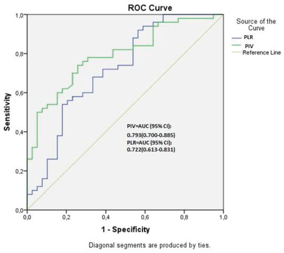
PLR: Platelet to lymphocyte ratio, PIV: Pan-immune-inflammation value, AUC: Area under the curve, CI: Confidence interval
DISCUSSION
In this study, inflammatory markers such as PIV, PLR, SII, LMR, SIRI, and NLR were detected at higher levels in patients with SVG disease. Among these inflammation markers, higher PIV and PLR better predicted the development of SVG disease. PIV includes more comprehensive blood parameters, which makes it a better predictor than PLR (AUC 0.793 vs. 0.722). This suggests that PIV may be a more accurate and comprehensive index in predicting immunological and inflammatory/anti-inflammatory conditions. These findings suggest that systemic inflammation plays a central role in the pathophysiology of SVG failure and that PIV could serve as a novel biomarker for assessing long-term graft patency. Today, CABG is the first option for left main coronary artery lesions and multivessel coronary artery diseases. However, the rapid progression of the atherothrombotic process in vein grafts compared to arterial grafts is the most important problem after CABG. Conditions such as hypertension, diabetes, and obesity cause damage to the SVG wall, resulting in increased expression and secretion of proinflammatory cytokines that promote the formation of atheromatous plaques. It is known that the use of arterial grafts increases the patency rate of the grafts and reduces the frequency of angina and re-intervention rates after CABG. Therefore, the use of the left internal mammary artery (LIMA) is the gold standard in left anterior descending artery revascularization. Venous grafts are easily accessible and do not spasm. However, it also has disadvantages such as having valves, limited adaptation to arterial pressure, diameter incompatibility, and rapidly progressing atherosclerosis. Early graft occlusion can be caused by a variety of factors, including the choice of grafts, the site of the anastomosis, severe graft stretching, and inflammation.
While LIMA has an 85% 10-year patency rate, SVG has a 61% rate. When we examine the mechanisms responsible for SVG occlusion, thrombosis in the first month after surgery, neointimal hyperplasia between 1-12 months, and atherosclerosis after the 12th month is at the forefront. Inflammation is known to be involved in all stages of atherosclerotic diseases. The process is mostly initiated by monocytes and lymphocytes secreting growth hormones and cytokines including platelet derived growth factor, interleukin-6, and interleukin-1. After entering the subendothelial layer to phagocytose oxidized low density lipoprotein particles, monocytes undergo a metamorphosis into foamy cells and contribute to the creation of the central region of atheroma plaque. High concentrations of monocytes and WBC have been related to an increased risk of coronary artery disease, according to Olivares et al.. Additionally, in cases of acute coronary syndrome, de LMR creased lymphocyte counts have been related to poor cardiovascular outcomes. We demonstrated, in our study, that two indicators that may be utilized to predict the patency of SVG are low levels of LMR and high levels of NLR. Supporting our study, previous studies have found that low LMR levels are associated with poor endpoints in both critical limb ischemia and in-stent restenosis, . PIV was related to long-term mortality in ST-elevation myocardial infarction, according to research by Murat et al. A relationship was found between PIV and no-reflow phenomenon in patients who underwent PCI after ST-segment elevation myocardial infarction (STEMI). In different studies, PIV has been found to be a good inflammatory index in predicting cardiovascular adverse events after STEMI and prognosis in heart failure, . To our knowledge, this is one of the few studies to investigate PIV in the context of late SVG patency. Our results complement prior studies, which have demonstrated associations between systemic inflammation and vein graft failure, though relying on simpler markers like CRP or WBC count, By contrast, PIV offers a more nuanced measure that reflects both innate and adaptive immune activity. This could explain its superior predictive value for SVG stenosis, as inflammation is involved in every phase of vein graft atherosclerosis from early thrombus formation to late plaque progression. PIV has also been associated with poor coronary collateral circulation. It has been demonstrated that some inflammatory markers are associated with major amputation in peripheral arterial diseases. Furthermore, our findings may have clinical implications. Identifying patients with elevated PIV or PLR prior to or after CABG could help in risk stratification, enabling more intensive monitoring, pharmacological intervention, or consideration of arterial graft alternatives. Statin therapy, for instance, has been shown to reduce inflammatory markers and improve SVG outcomes.
In the present study, we investigated many indexes that may be used to predict late SVG occlusion. In Multivariate logistic regression analysis, we found that PLR and PIV were independent predictors of SVG. Whether PIV-guided anti-inflammatory strategies can further enhance graft patency warrants prospective evaluation.
Study Limitations
This study has several limitations that should be acknowledged. First, it is a single-center, retrospective study, which may limit the generalizability of the results to broader populations. We were unable to obtain detailed data on statin use and smoking. Second, although we attempted to exclude patients with active infections, malignancies, or chronic inflammatory conditions, subclinical inflammation or undiagnosed conditions may have influenced systemic inflammatory markers. Third, the observational nature of the study precludes establishing a causal relationship between inflammatory markers such as PIV and PLR and SVG disease. Fourth, the study population size, while adequate for preliminary analysis, may still be insufficient to detect small but clinically significant associations. Additionally, the inflammatory markers were measured only once before angiography; serial measurements over time could provide a more dynamic and accurate understanding of the inflammatory state. Although angiographic assessment was performed by two blinded cardiologists, visual estimation of stenosis can still be subject to interobserver variability, and no intravascular imaging modalities were used to confirm graft patency or characterize plaque morphology. Another limitation of our study is that it is not precisely stated when saphenous graft disease occurs since these patients were not followed regularly after CABG surgery.
CONCLUSION
We showed in this study that two of the novel inflammatory indexes, PIV and PLR, may be significant predictors of saphenous graft patency following coronary by-pass surgery. SVG disease can be predicted from routine blood tests that can be calculated simply and practically. In clinical practice, these indexes may help specialists in predicting saphenous graft stenosis in patients with CABG.
Footnotes
Ethics
Ethics Committee Approval: The study was authorized by Tekirdağ Namık Kemal University Local Ethics Committee (decision no: 2021.284.12.07, date: 28.12.2021) and was carried out by the Helsinki Declaration.
Informed Consent: It is a single-center, retrospective study.
Authorship Contributions
Surgical and Medical Practices: C.A., Concept: A.D., Design: H.O., Data Collection or Processing: C.A., Analysis or Interpretation: A.D., Literature Search: H.O., Writing: C.A.
Conflict of Interest: No conflict of interest was declared by the authors.
Financial Disclosure: The authors declared that this study received no financial support.
References
Nwaejike N, Tennyson C, Mosca R, Venkateswaran R. Reusing the patent internal mammary artery as a conduit in redo coronary artery bypass surgery. Interact Cardiovasc Thorac Surg. 2016;22:346-50.
Ross R. Atherosclerosis--an inflammatory disease. N Engl J Med. 1999;340:115-26.
Karadeniz M, Aydın C, Demirkıran A, Alp Ç. Predictors of severity of coronary artery disease in patients with acute st-elevation myocardial ınfarction. Namik Kemal Med J. 2024;12:171-5.
Bagger JP, Zindrou D, Taylor KM. Leukocyte count: a risk factor for coronary artery bypass graft mortality. Am J Med. 2003;115:660-3.
Turak O, Ozcan F, Isleyen A, Tok D, Sokmen E, Buyukkaya E, et al. Usefulness of the neutrophil-to-lymphocyte ratio to predict bare-metal stent restenosis. Am J Cardiol. 2012;110:1405-10.
Aydın C, Uyan U, Karadeniz M, Demirkıran A. Role of simple inflammatory parameters in predicting the severity of coronary artery disease. Rev Assoc Med Bras (1992). 2023;69:e20230518.
Kurtul S, Sarli B, Baktir AO, Demirbas M, Saglam H, Doğan Y, et al. Neutrophil to lymphocyte ratio predicts SYNTAX score in patients with non-ST segment elevation myocardial infarction. Int Heart J. 2015;56:18-21.
Goldman S, Zadina K, Moritz T, Ovitt T, Sethi G, Copeland JG, et al. Long-term patency of saphenous vein and left internal mammary artery grafts after coronary artery bypass surgery: results from a department of veterans affairs cooperative study. J Am Coll Cardiol. 2004;44:2149-56.
Parang P, Arora R. Coronary vein graft disease: pathogenesis and prevention. Can J Cardiol. 2009;25:e57-62.
Oude Nijhuis MM, van Keulen JK, Pasterkamp G, Quax PH, de Kleijn DP. Activation of the innate immune system in atherosclerotic disease. Curr Pharm Des. 2007;13:983-94.
Olivares R, Ducimetière P, Claude JR. Monocyte count: a risk factor for coronary heart disease? Am J Epidemiol. 1993;137:49-53.
Bian C, Wu Y, Shi Y, Xu G, Wang J, Xiang M, et al. Predictive value of the relative lymphocyte count in coronary heart disease. Heart Vessels. 2010;25:469-73.
Gary T, Pichler M, Belaj K, Eller P, Hafner F, Gerger A, et al. Lymphocyte-to-monocyte ratio: a novel marker for critical limb ischemia in PAOD patients. Int J Clin Pract. 2014;68:1483-7.
Murat SN, Yarlioglues M, Celik IE, Kurtul A, Duran M, Kilic A, et al. The relationship between lymphocyte-to-monocyte ratio and bare-metal stent ın-stent restenosis in patients with stable coronary artery disease. Clin Appl Thromb Hemost. 2017;23:235-40.
Murat B, Murat S, Ozgeyik M, Bilgin M. Comparison of pan-immune-inflammation value with other inflammation markers of long-term survival after ST-segment elevation myocardial infarction. Eur J Clin Invest. 2023;53:e13872.
Bayramoğlu A, Hidayet Ş. Association between pan-immune-inflammation value and no-reflow in patients with ST elevation myocardial infarction undergoing percutaneous coronary intervention. Scand J Clin Lab Invest. 2023;83:384-9.
Liu Y, Liu J, Liu L, Cao S, Jin T, Chen L, et al. Association of systemic ınflammatory response ındex and pan-ımmune-ınflammation-value with long-term adverse cardiovascular events in ST-segment elevation myocardial ınfarction patients after primary percutaneous coronary ıntervention. J Inflamm Res. 2023;16:3437-54.
Inan D, Erdogan A, Pay L, Genc D, Demırtola AI, Yıldız U, et al. The prognostic impact of inflammation in patients with decompensated acute heart failure, as assessed using the pan-immune inflammation value (PIV). Scand J Clin Lab Invest. 2023;83:371-8.
Yayla C, Gayretli Yayla K. C-Reactive protein to albumin ratio in patients with saphenous vein graft disease. angiology. 2021;72:770-5.
Yavuz S, Engin M, Yazgan E, Demir OF, Turk T. Letter: potential predictors of saphenous vein graft disease after coronary artery bypass operations. Angiology. 2022;73:689-90.
Yilmaz Y, Kelesoglu S. The Importance of Pan-immune inflammation value (PIV) in predicting coronary collateral circulation in stable coronary artery patients. Angiology. 2024:33197241258529.
Çakmak AC, Vatan MB, Sarıbıyık Çakmak B, Erkin A, Tatlı E, Kocayigit I. The systemic immune-inflammation index is an independent predictive factor in predicting major amputation in chronic limb-threatening ischemia. Vascular. 2025;0
Serruys PW, de Feyter P, Macaya C, Kokott N, Puel J, Vrolix M, et al. Fluvastatin for prevention of cardiac events following successful first percutaneous coronary intervention: a randomized controlled trial. JAMA. 2002;287:3215-22.